EN


How to Evaluate X-Ray Machine Generator Power and Image Quality
When a hospital compares an x ray machine, generator power is often the first specification discussed. It matters, but it is not a stand-alone measure of image quality. A useful evaluation connects the generator to the examinations the department performs, the detector and image-processing workflow, and the quality-control process that will verify performance after installation.
From a medical-equipment selection perspective, the practical question is not simply “How many kilowatts?” It is whether the x ray machine can produce the required exposure factors consistently for the intended workload while the imaging chain delivers usable detail, contrast, and noise performance at an appropriate exposure. That distinction helps procurement, radiography, biomedical engineering, and facilities teams compare proposals on the same basis.
Start With the Clinical Workload, Not a Power Number
Generator requirements should follow the planned examination mix. A room used mainly for routine adult chest, extremity, and basic table examinations has a different demand profile from one supporting heavier patients, trauma, mobile work, or frequent high-throughput studies. Before reviewing a quotation for a medical X-ray machine, document the patient population, projections, detector sizes, expected examinations per shift, room workflow, and whether automated exposure control is part of the configuration.
Generator ratings are commonly stated in kilowatts, but the rating represents available electrical output under specified operating conditions. It does not by itself tell a team which techniques can be used in every situation. Ask the supplier to show the relevant kV, mA, mAs, exposure-time ranges, and the combinations available at clinically relevant stations. In other words, compare the usable exposure envelope, not a headline power rating alone.
- kV: influences beam energy and subject contrast; it must suit the anatomy and the department’s technique charts.
- mA and exposure time: affect how quickly the required mAs can be delivered. Shorter exposures may be important where motion is a concern.
- mAs range and accuracy: determine whether routine and more demanding techniques can be selected reproducibly.
- Heat loading and duty cycle: show whether the tube-generator combination fits the anticipated sequence of examinations rather than only an isolated exposure.
If the target department’s workflow and configuration conditions are the same, this comparison is more meaningful. A higher rating may provide operational headroom, but it is not automatically the right choice for every x ray machine installation.
Translate Generator Specifications Into Real Examination Requirements
Build a requirements-to-configuration table before scoring systems. List representative adult and pediatric projections, expected patient-size range, desired exposure-time limits, and expected room throughput. Then require each bidder to map those tasks to the proposed diagnostic radiography equipment configuration. This makes it easier to identify a mismatch between a nominal generator rating and the techniques a department actually needs.
| Evaluation question | What to request | Why it matters |
|---|---|---|
| Can the system support the planned technique chart? | Available kV/mA/mAs combinations and exposure-time limits | Confirms that output is usable for intended projections. |
| Will performance remain stable through a busy session? | Tube heat-unit information, cooling behavior, and stated duty conditions | Connects the generator and tube to throughput expectations. |
| How is exposure controlled? | AEC chamber configuration, backup settings, and setup guidance | Shows how the system supports repeatable exposure selection. |
| How will acceptance be verified? | Commissioning tests, baseline measurements, and service documentation | Turns specifications into observable performance checks. |
Do not ask only whether a system has an automatic feature. Ask how it will be verified under the target patient-equivalent, detector, grid, and technique conditions. This is particularly important when a digital radiography system is expected to support several rooms or a broad range of operators.
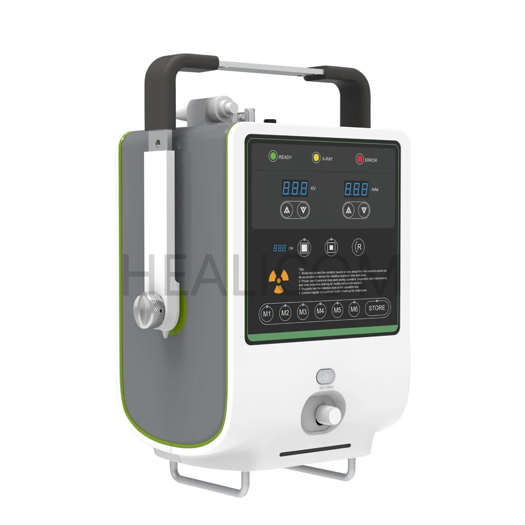
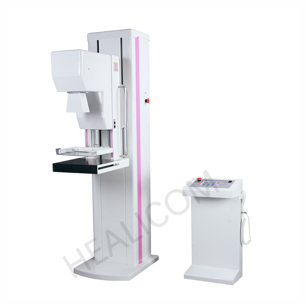 Evaluate generator capability as part of the complete radiography workflow.
Why Generator Power Does Not Equal Image Quality
An image is produced by a chain, not by the generator alone. The generator supplies the selected exposure; the tube and filtration shape the beam; the patient and positioning affect attenuation; the detector captures signal; and processing, display, and viewing conditions influence what users see. A capable generator can still be paired with a workflow that produces avoidable noise, saturation, artifacts, or inconsistent appearance.
The International Atomic Energy Agency describes radiography optimization as balancing contrast, detail, and noise with patient exposure. That is a helpful procurement principle: evaluate whether the complete imaging chain can achieve the department’s clinical task at the exposure level set by local protocols. More output is not a substitute for correct technique, detector calibration, collimation, positioning, or quality assurance.
For a hospital imaging equipment project, separate these questions: can the generator deliver the required technique in time; does the detector suit the intended exams; does processing preserve useful information; and can the department establish baselines after commissioning? The priority is not the number of listed functions, but whether they are needed and can be checked in the target workflow.
Assess Image Quality With a Repeatable Test Plan
Ask for an acceptance and quality-control plan before selecting an x ray machine. The plan should name the test objects or phantoms, baseline images, exposure settings, image-review criteria, measurement tools, responsible roles, and corrective path. Where applicable, confirm how the plan relates to local regulation, the manufacturer’s instructions, and the department’s medical physics program.
Key image-quality dimensions
- Spatial detail: assess whether fine structures needed for the clinical task are visible under controlled test conditions.
- Contrast and low-contrast visibility: review whether processing and exposure settings distinguish relevant differences without obscuring anatomy.
- Noise: compare noise appearance and signal consistency at representative techniques rather than relying on a single demonstration image.
- Uniformity and artifacts: inspect detector response, stitching where used, grids, collimation, and processing for repeatable issues.
- Exposure indicators: agree how exposure index or related feedback will be monitored so staff can identify technique drift.
Quality-control guidance for diagnostic radiology uses baseline measurements and tolerances to make changes visible. The precise values must be set by the applicable protocol and responsible physics team. Treat an exposure indicator as a process signal, not a universal pass/fail diagnosis of image quality.
Compare a Digital Radiography System Beyond the Detector Datasheet
In a digital radiography system, detector specifications are important, but integration determines whether those specifications translate into routine use. Review detector availability, charging and battery workflow for wireless units, image-transfer reliability, workstation ergonomics, image-processing presets, exposure-index display, DICOM connectivity, and backup procedures. A technically capable detector can create delays or inconsistency if its workflow is poorly aligned with the room.
Request a task-based demonstration. For example, have the supplier show the planned chest workflow from patient setup through image acquisition, repeat analysis, routing, and review. Then repeat with a representative high-attenuation or mobility-constrained scenario. The goal is not to force a sales demonstration into a clinical claim; it is to observe how the proposed configuration supports the actual process.
Use a Procurement Scorecard That Links Power to Verification
A practical scorecard prevents one attractive specification from dominating a decision. Weight categories according to the department’s documented priorities, and require evidence for every score.
- Clinical coverage: supported projections, patient range, and workflow constraints.
- Generator and tube capability: usable exposure envelope, timing, heat management, and repeatability documentation.
- Image chain: detector, processing, display, and integration checks.
- Safety and compliance review: documentation for the applicable market and standards; for projection radiography, IEC 60601-2-54 is one technical framework to discuss with qualified reviewers.
- Commissioning and service: acceptance tests, baseline records, training, maintenance responsibilities, and escalation path.
- Lifecycle fit: installation conditions, electrical and space requirements, uptime planning, and maintenance resources.
When evaluating an x ray machine, move from parameter-first to workflow-first thinking: put the proposed system into the examination process, then see whether its technical limits support each step. Also move from a function list to verification questions: what measurement, image review, or acceptance record will demonstrate that the feature works as intended?
Questions to Resolve Before Purchase Approval
- Which examination types and patient conditions define the required exposure envelope?
- Which generator settings can be achieved at the desired exposure times and duty conditions?
- What are the detector, processing, and display limitations that may affect image quality?
- Which acceptance tests will establish baseline output, image quality, and exposure indicators?
- Who will own quality-control review, service coordination, and protocol changes after go-live?
- Which site conditions—power supply, room shielding, network, space, and staffing—must be confirmed before installation?
Conclusion
A sound x ray machine evaluation does not treat generator power as a shortcut for image quality. Match the generator’s usable output to real examination requirements, examine the entire image chain, and require a commissioning plan with measurable baselines. That approach gives a procurement team a clearer way to compare a medical X-ray machine, diagnostic radiography equipment, and a digital radiography system while keeping clinical workflow and verification at the center of the decision.


 Hot News