EN


What Quality Assurance Tests Are Important for X-Ray Machines?
Quality assurance for an x ray machine is not one pass-or-fail event. It is a documented program that confirms whether the imaging chain continues to perform as the facility expects: from the generator and exposure controls through the detector, display, records, and safety devices. For a medical X-ray machine, the local program should be written and reviewed by qualified personnel, including a medical physicist where required by the facility and jurisdiction.
A practical program starts with a baseline. Acceptance tests establish what the x ray machine can do when it is installed or returned to service. Routine tests then look for meaningful drift before image quality, workflow, or radiation management is affected. The exact test methods, tolerances, and frequencies belong in the site protocol; a checklist is useful only when it reflects the particular diagnostic radiography equipment, workload, and applicable regulations.
Start with acceptance and baseline testing
Before clinical use, document the configuration, detector serial information, software version, exposure techniques, accessories, and room conditions for the x ray machine. The acceptance record gives the team a defensible reference for later comparisons. A qualified test plan normally checks that the delivered system matches the agreed configuration and that its measured performance is suitable for the intended examinations.
For a digital radiography system, baseline images should be acquired with the site’s approved phantoms and routine protocols. Retain representative images, exposure indices, and test settings. If a later result changes, the team can compare like with like rather than relying on a visual memory of how the x ray machine used to perform.
Generator and exposure-control tests
Generator checks examine whether the x ray machine produces exposures consistently and as selected. Common items include:
- kVp accuracy and repeatability: compare selected and measured tube potential across relevant technique ranges. A shift may alter beam quality, contrast behavior, and dose planning.
- Exposure-time accuracy: verify short and long exposure times used by the department. Timing matters especially where motion control or automatic exposure termination is involved.
- Output reproducibility and linearity: repeat fixed exposures and compare output as technique settings change. These checks help reveal instability in the generator or tube circuit.
- Automatic exposure control performance: assess repeatability, chamber selection, backup timing, and response to representative attenuation. AEC results should be interpreted with the protocol, receptor, and phantom setup clearly recorded.
These tests are not interchangeable. A stable-looking image does not by itself show that generator output is stable, and an acceptable output measurement does not replace a detector review. For hospital imaging equipment, trend data is often more useful than a single isolated measurement.
Beam quality, collimation, and dose-related checks
Beam quality and geometry checks connect technical output to safe, repeatable positioning. The QA plan for an x ray machine commonly includes total filtration or half-value layer verification where applicable, beam alignment, light-field to radiation-field congruence, and collimator accuracy. The team may also review source-to-image receptor distance indicators, locks, detents, and tube-stand movement.
For mobile or room-based diagnostic radiography equipment, test the functions that staff depend on during setup: exposure switches, interlocks, warning indicators, emergency stops, and mechanical supports. Any abnormal noise, drift, damaged cable, failed lock, or unreliable indicator should be recorded and assessed rather than normalized as part of daily work.
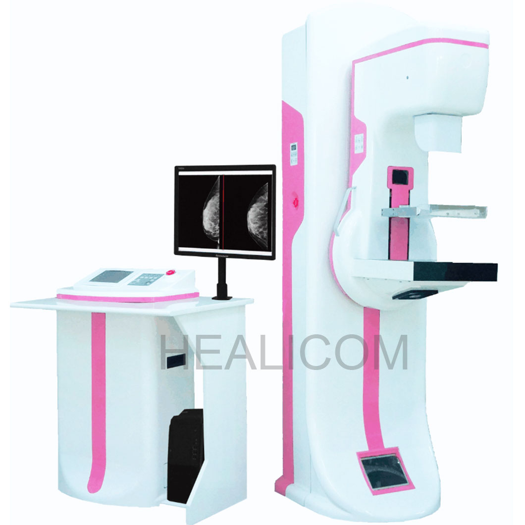 A planned QA review combines phantom imaging, measurement, and documented comparison with baseline results.
Image receptor and digital image-quality tests
A digital radiography system needs QA beyond the generator. Detector-focused testing may include uniformity, bad-pixel or artifact review, exposure-index consistency, signal response, residual image checks, and contrast-detail performance with a suitable phantom. The goal is not simply to make the image brighter or darker; it is to confirm that the receptor, processing pathway, and display workflow are behaving predictably.
When evaluating an x ray machine, inspect images at the workstation used in practice. A defect that is invisible on one display can still affect interpretation elsewhere. Record the protocol, processing settings, monitor conditions, and any rejected-image pattern. Repeated positioning or exposure errors can point to training, workflow, or user-interaction issues rather than an equipment fault alone.
Mechanical and safety tests
Mechanical reliability is part of image quality and safety. Test table movement, wall stand travel, tube suspension, brakes, detents, compression components where fitted, and collision-prevention features according to the equipment type. Verify that labels, warning lights, audible signals, and protective devices remain legible and functional. A medical X-ray machine should also be checked after relocation, detector replacement, major software change, repair, or an incident that could affect calibration.
Set a frequency that matches the risk
Daily or pre-use checks are usually simple observations: inspect cables, controls, locks, detector condition, warning indicators, and a test image if the local protocol calls for one. More detailed checks may be weekly, monthly, quarterly, annually, or triggered by service events. The right interval for an x ray machine depends on modality, usage, manufacturer instructions, local regulation, and medical-physics policy. Do not copy a frequency from another site without confirming that it applies to your configuration.
Turn QA results into a usable record
Every test record should identify the x ray machine, date, operator, test equipment, phantom, technique factors, measured result, baseline or tolerance, and disposition. Define who reviews failures, who can remove equipment from clinical use, and how retesting is documented after service. A concise trend chart or controlled log can make gradual changes visible before they become a complaint or repeat-exposure issue.
For procurement and planning, ask suppliers how their hospital imaging equipment supports acceptance documentation, service records, detector QA, and training. That discussion is more useful than comparing a feature list alone. It helps the facility determine whether the medical X-ray machine and its support process can fit its actual quality program.
A practical QA checklist
- Establish acceptance baselines for the x ray machine before clinical handover.
- Test generator accuracy, output reproducibility, timing, and AEC with approved methods.
- Check beam alignment, collimation, mechanical controls, and safety features.
- Evaluate detector uniformity, artifacts, exposure-index behavior, and image quality on the clinical workflow.
- Document trends, investigate deviations, and repeat relevant tests after service or configuration changes.
In short, the important QA tests are the ones that link the performance of an x ray machine to its intended clinical use. A qualified, site-specific program should cover generator output, beam geometry, receptor performance, image review, mechanical safety, and documented follow-up. That combination gives the imaging team a clearer basis for deciding when the x ray machine is ready for use and when it needs further evaluation.

 Hot News