EN


What Is an X-Ray Machine and How Is It Used in Medical Imaging?
An x ray machine is a medical imaging system that directs a controlled X-ray beam through part of the body and records the differing amounts of radiation that reach a detector. Dense structures such as bone usually attenuate more of the beam than air-filled tissue, creating contrast that trained clinicians can interpret. In everyday practice, an x ray machine is not just a tube and a screen: it is a coordinated imaging chain that includes the generator, X-ray tube, patient-positioning arrangement, detector, image-processing software, display, and local safety procedures.
For a hospital team, understanding how an x ray machine works makes it easier to separate useful system questions from a long specification sheet. It also helps purchasing, radiology, and facilities staff agree on what must be verified before installation and acceptance. The following overview is educational and does not replace local clinical protocols, radiation-safety requirements, or qualified professional judgment.
How an X-Ray Machine Produces an Image
At the start of an exposure, the generator supplies electrical energy to the X-ray tube. Inside the tube, electrons are accelerated toward a target; their interaction produces X-rays. Collimation shapes the beam to the anatomy being examined, while positioning and exposure settings affect the image that the detector receives. The detector turns the remaining radiation into image information for review.
A modern medical X-ray machine commonly uses a digital detector, which is why the term digital radiography system is often used in discussions of current workflows. The detector and software can present the image rapidly, but speed is only one part of the task. The x ray machine still depends on appropriate positioning, exposure selection, image review, and repeat-exposure control. A digital image that looks acceptable on a display does not by itself prove that the examination met every local quality or clinical requirement.
The Main Parts of an X-Ray Machine
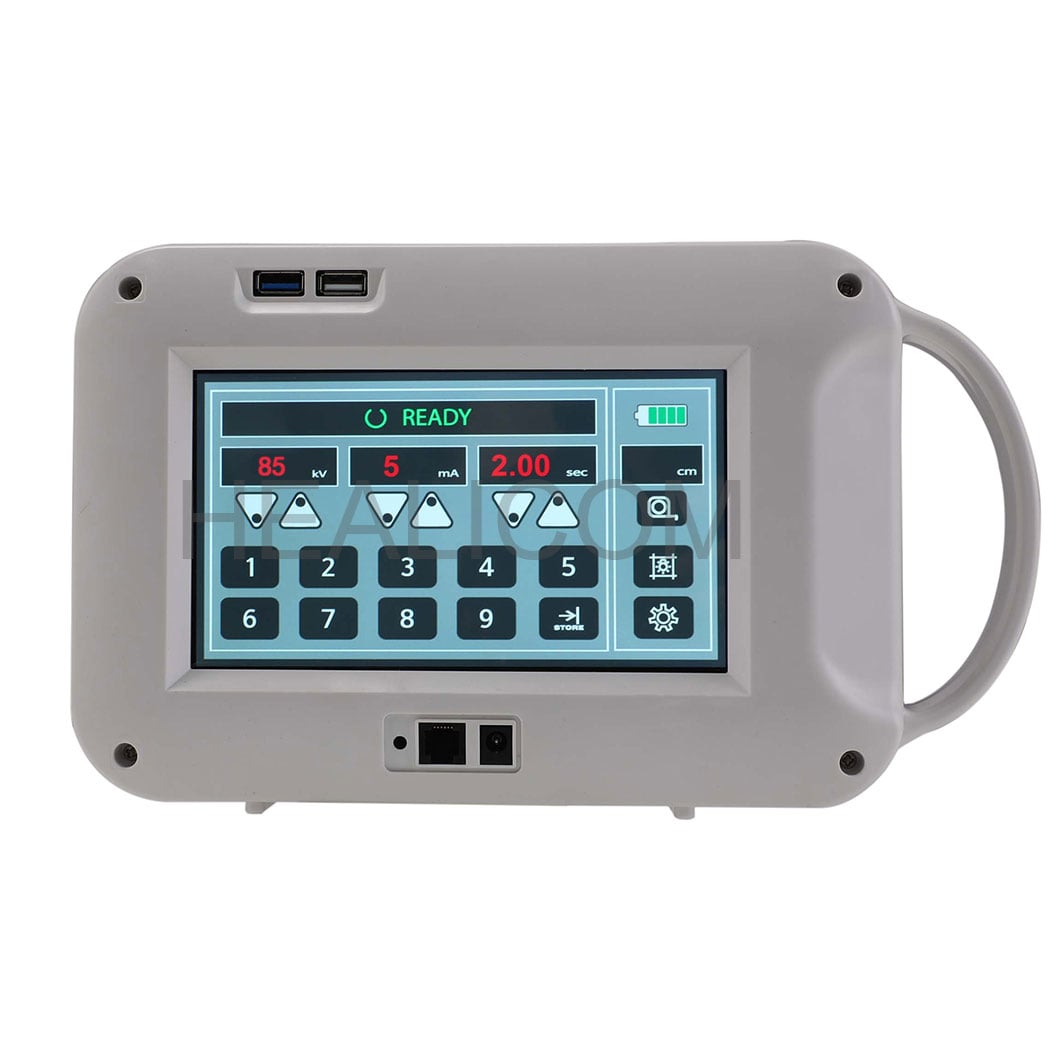
- Generator and control console: The generator supplies controlled electrical power. The console lets authorized operators select exposure factors within the facility’s protocol.
- X-ray tube and collimator: The tube creates X-rays and the collimator limits the beam to the region of interest. Beam restriction is a practical safety and image-quality consideration.
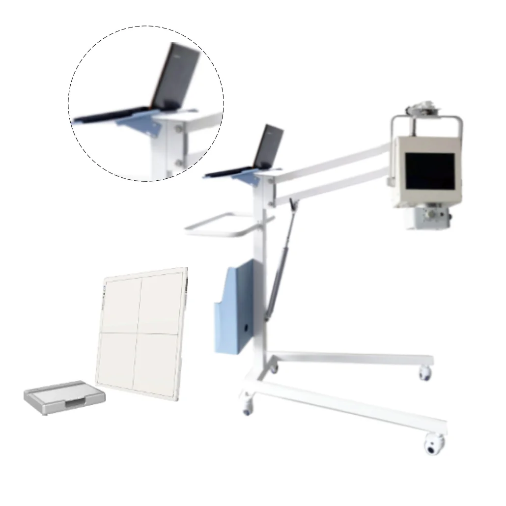
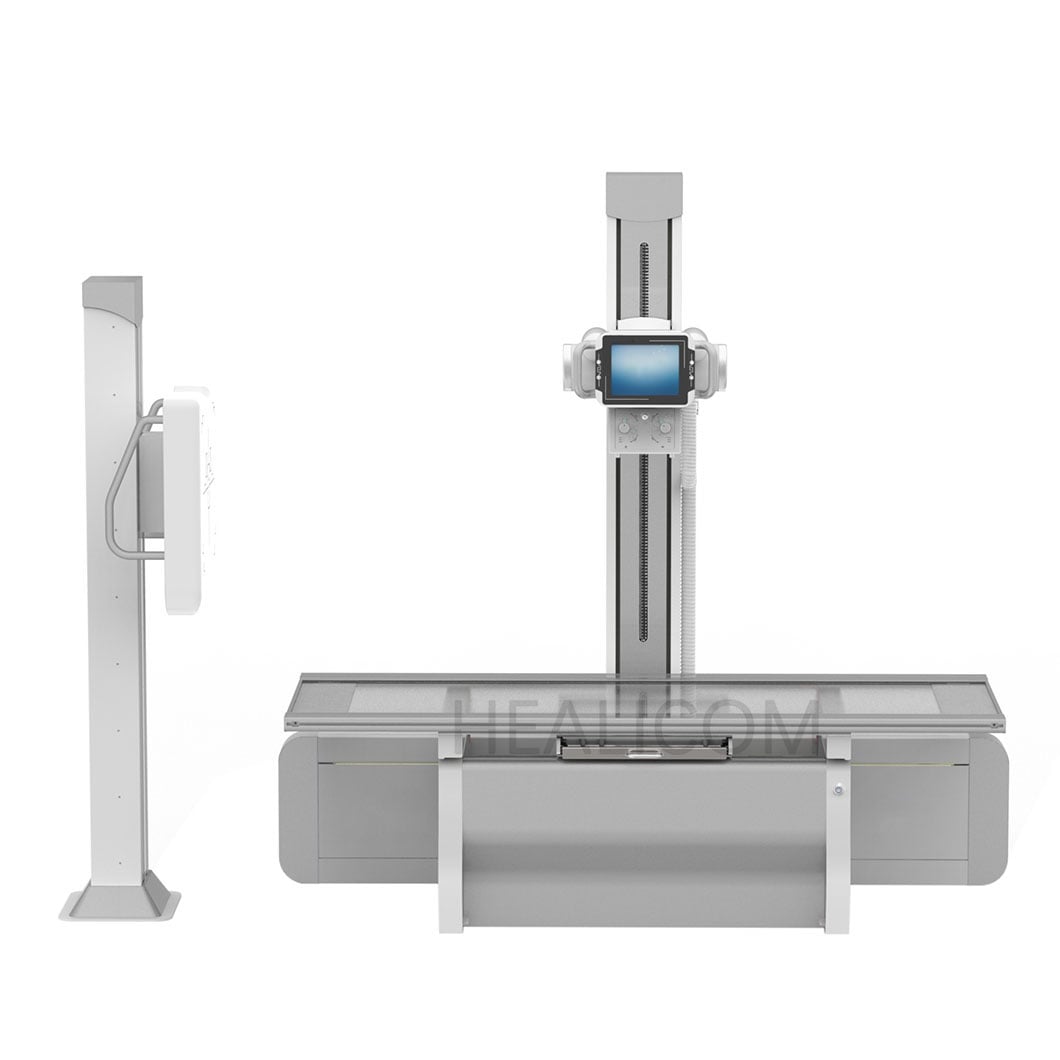
- Patient support and positioning accessories: A table, wall stand, bucky, or mobile arrangement helps place the patient and detector consistently for the intended examination.
- Detector and processing chain: In a digital radiography system, the detector captures image data and software processes it for review, storage, and transfer according to the facility workflow.
- Display, storage, and connectivity: The x ray machine may connect to a local imaging environment. Compatibility, workflow mapping, and cybersecurity should be checked with the relevant technical teams rather than assumed.
Common Types of Diagnostic Radiography Equipment
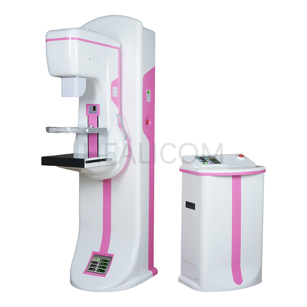
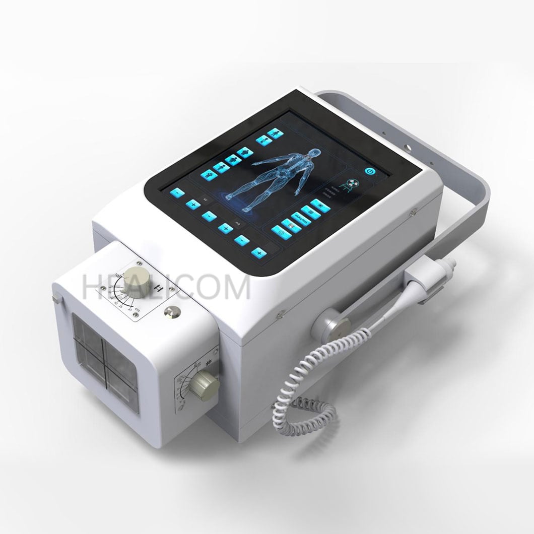
Diagnostic radiography equipment is configured around clinical setting, patient mobility, examination mix, available space, and local workflow. A fixed room-based x ray machine may suit sites with a predictable examination volume and a planned radiography room. Mobile systems support imaging where moving the patient is difficult, but they also require clear routes, storage, charging or power planning, and bedside workflow controls. Compact or portable formats can address particular use cases; they should be evaluated against the same questions about operator training, intended use, image workflow, and applicable regulation.
Another decision is the imaging chain. Some facilities are updating older computed-radiography workflows, while others consider a direct digital radiography system. Rather than treating detector technology as an automatic answer, compare it against the actual task: turnaround expectations, existing image-management environment, service coverage, staff familiarity, and the examinations the site performs. A feature only has value when it fits the daily process it is meant to support.
How X-Ray Machines Are Used in Medical Imaging
A clinician may request an x ray machine examination to visualize bones, chest structures, joints, or other anatomy when radiography is an appropriate part of the diagnostic pathway. The acquisition team follows the facility’s procedure for identification, positioning, exposure, review, documentation, and escalation. The image is then interpreted by appropriately qualified professionals in the local care setting.
This division of responsibilities matters. The x ray machine creates image data; it does not make a diagnosis by itself. Likewise, a purchasing comparison should not turn a list of generator, detector, or software features into a promise about patient outcomes. When a team compares hospital imaging equipment, the better question is whether each component supports the required examinations, staffing pattern, room conditions, and quality-control plan.
Safety, Training, and Quality Control
Because an x ray machine uses ionizing radiation, implementation includes more than delivery and installation. U.S. FDA materials describe radiation-safety performance standards for X-ray imaging systems and emphasize appropriate credentials and radiation-safety training for personnel. Requirements vary by country, intended use, and local authority, so facilities should confirm the rules that apply to their own project.
Quality assurance is equally practical. The International Atomic Energy Agency identifies performance monitoring and quality control as important for accurate and safe diagnostic radiology use. For an x ray machine, a workable plan can define acceptance checks, routine tests, image-review responsibilities, service contacts, fault reporting, and recordkeeping. Change the lens for a moment: the real procurement question is often how the team will verify performance after installation, not merely which feature appears on a brochure.
A Practical Evaluation Checklist for Hospital Imaging Equipment
When comparing an x ray machine, assess the project in three stages: requirements, configuration, and acceptance. That structure helps avoid a common mistake—assuming that a listed specification automatically fits a department’s workflow.
- Define the examination and workflow scope. Identify the patient population, procedure types, anticipated volume, mobility needs, and who will operate the system.
- Confirm the room and infrastructure. Check space, shielding design, electrical supply, network readiness, access routes, and any local construction or approval requirements.
- Review the configuration in context. Compare the detector format, positioning arrangement, console workflow, image routing, and available accessories against documented use cases.
- Plan training and quality control. Clarify operator training, acceptance testing, periodic checks, maintenance response, and escalation contacts before commissioning.
- Document handover evidence. Keep records for installed configuration, training, test results, and service responsibilities so the x ray machine can be managed over its operating life.
Healicom Medical Equipment Co.,LTD. covers imaging equipment alongside other medical-equipment categories. Teams beginning a broader sourcing discussion can review the company’s medical equipment resources, then confirm the configuration and local compliance needs for their specific project.
Key Takeaway
An x ray machine is a complete radiography system, not a single component. It produces image data by combining controlled X-ray generation, patient positioning, detection, processing, and professional review. Whether the project involves a fixed room, a mobile unit, or a digital radiography system, the soundest evaluation links equipment choices to clinical workflow, room readiness, trained use, quality control, and local safety requirements. That condition-based approach gives a hospital team a clearer basis for selecting and managing diagnostic radiography equipment.

 Hot News