EN


How to Compare CBCT Field of View and Image Resolution
When a dental team compares a CBCT system, field of view and image resolution should be evaluated together rather than as separate specifications. A scan that covers more anatomy may be appropriate for one task, while a smaller volume and finer voxel setting may be more useful for another. The practical question is not “Which setting has the highest number?” but “What anatomy and diagnostic task must this scan support?”
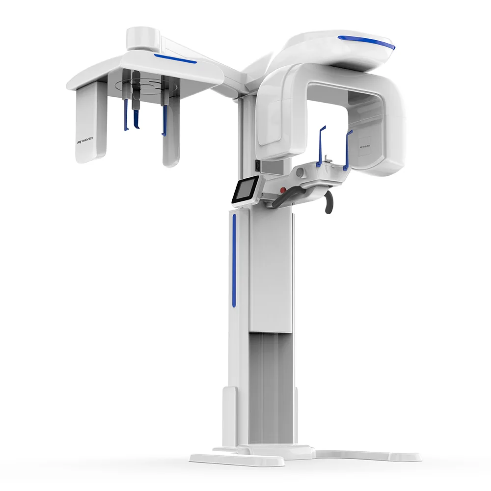
From a biomedical engineering perspective, the most useful comparison starts with clinical workflow, intended anatomy, and review requirements. A dental CBCT scanner should be assessed as a protocol system: field of view, voxel size, exposure settings, patient positioning, reconstruction, and display conditions all shape the image that the clinician finally reviews.
Start with the difference between field of view and resolution
In CBCT, the field of view (FOV) is the physical volume captured in a scan. It is commonly described by height and diameter or by a named category such as limited, medium, or large FOV. Image resolution describes the system’s ability to represent fine spatial detail. Voxel size is an important part of that discussion: a voxel is the three-dimensional counterpart of a pixel, and smaller voxels can support representation of smaller structures when the full scan protocol and system performance are suitable.
These terms are related but not interchangeable. A small FOV does not automatically create a diagnostically useful image, and a small voxel number does not by itself establish image quality. Noise, contrast, motion, artifacts, reconstruction settings, and the anatomy’s position inside the volume also matter. For a cone beam CT system comparison, ask suppliers to show the complete protocol rather than quoting one resolution number in isolation.
Match the CBCT field of view to the clinical question
A sensible CBCT field of view begins with the smallest volume that includes the anatomy required for the justified examination. This approach can reduce unnecessary coverage and gives the team a clearer basis for protocol selection. The exact FOV labels and dimensions differ between systems, so compare actual scan dimensions rather than relying only on labels such as “small” or “medium.”
| Typical task | FOV decision point | Resolution question to ask |
|---|---|---|
| Localized endodontic assessment | Can a limited volume include the tooth, root area, and relevant adjacent anatomy? | Does the protocol offer a voxel setting appropriate for the fine detail being evaluated? |
| Implant planning for a local site | Does the selected volume show the proposed site and relevant anatomical boundaries? | Is the image sufficiently clear to support the planned measurements and review process? |
| Single- or dual-arch assessment | Is a medium CBCT field of view needed to capture both the area of interest and the required reference anatomy? | Which voxel and exposure options are available at that volume? |
| Orthodontic or broader maxillofacial review | Is a larger volume justified by the anatomy and treatment-planning question? | What tradeoffs in detail, artifact control, and review workflow arise at the larger FOV? |
For example, an approximately 8 × 8 cm volume is often described in educational materials as a medium FOV for single- or dual-arch work, but it is only an illustrative reference. The correct choice depends on anatomy, patient size, clinical purpose, and the system’s available protocols.
How voxel size affects a 3D dental scan
Voxel size is usually expressed in micrometers or millimeters. A smaller voxel can help depict finer spatial detail, which is why it is frequently discussed for focused applications. Yet a 3D dental scan should not be selected by voxel size alone. Smaller voxels may require changes in acquisition conditions, can increase noise under some conditions, and can create larger image files and longer reconstruction or review demands.
Use a task-based comparison instead. If the team is evaluating a narrow anatomical question, ask for representative images from a limited-volume protocol and verify that the image can be reviewed effectively on the intended workstation. If the task requires a wider anatomical overview, confirm what image quality is available at the larger volume and whether the protocol still supports the information needed. Higher specification figures are not automatically better; the relevant issue is whether the CBCT protocol serves the target diagnostic task.
Compare image quality beyond the resolution specification
Resolution is only one component of usable image quality. During a dental CBCT scanner evaluation, include these questions in the comparison sheet:
- Spatial detail: Which voxel sizes and reconstruction options are available for each FOV?
- Contrast and noise: How do the available protocols perform when the team reviews the intended anatomy?
- Artifacts: How are metal restorations, patient movement, and dense structures managed in the displayed image?
- Positioning: Can the patient be positioned consistently so the region of interest sits correctly in the scan volume?
- Workflow: Who selects the protocol, reviews the scan, exports images, and confirms that the right volume was acquired?
- Verification: Can the supplier document the settings used for demonstration images and support site acceptance checks?
Do not ask only whether a system has a high-resolution mode. Ask how that mode changes across FOV sizes and whether it is available for the clinical scenarios your team actually performs. This turns a generic equipment comparison into a usable CBCT protocol review.
A practical comparison sequence for buyers
- List the intended examinations. Separate focused tooth-level tasks, local implant assessments, arch-level reviews, and broader craniofacial indications.
- Map each task to required anatomy. Define what must be inside the CBCT field of view, including relevant adjacent structures.
- Request a protocol matrix. For every FOV, record voxel options, exposure controls, scan time, reconstruction behavior, and file-handling requirements.
- Review representative images. Use de-identified demonstration cases or an agreed acceptance process that reflects the intended task. Do not treat marketing images as a substitute for verification.
- Check implementation conditions. Confirm positioning support, staff training, workstation performance, data storage, maintenance responsibilities, and local regulatory or clinical requirements.
- Document acceptance criteria. Agree on what will be checked after installation so the selected cone beam CT system can be assessed against the planned workflow.
A common purchasing mistake is treating a specification sheet as proof of clinical fit. It is more reliable to define the required FOV and image-quality conditions first, then compare which CBCT protocols meet them. This is especially important when one device offers several scan volumes and resolution settings.
Questions to take to a CBCT supplier
- Which FOV dimensions are available, and which protocol settings are tied to each volume?
- What voxel sizes can be selected for each FOV, and what are the practical limits or tradeoffs?
- Can the team review sample images for the intended local, arch-level, or broader task?
- How are positioning, motion, and metal artifacts addressed during normal use?
- What training and documentation support protocol selection and routine quality checks?
These questions help buyers compare a dental CBCT scanner on usable imaging workflow rather than on a single headline number. They also create a clearer discussion with suppliers about configuration scope, demonstration conditions, and post-installation verification.
Make the choice clinically appropriate and operationally workable
Comparing CBCT field of view and image resolution is a balancing exercise. The goal is to capture the required anatomy with a protocol that supports the intended clinical review, while avoiding an assumption that a larger volume or smaller voxel is inherently the right answer. Start with the clinical question, define the anatomy that must be included, compare the protocol options for that FOV, and document how the team will verify image usability after deployment.
This article offers an equipment-evaluation framework and does not replace clinical judgment, radiation-protection requirements, local regulations, or manufacturer instructions. When a facility is preparing a purchase or protocol update, a structured comparison table can help align clinicians, imaging staff, and procurement teams before a final CBCT decision.
 Hot News