Xray machines have been a cornerstone of medical diagnosis for over a century, helping doctors see inside the human body without invasive procedures. From broken bones and lung infections to dental cavities and internal tumors, an xray machine provides clear, detailed images that guide treatment decisions. But how exactly does this device turn invisible radiation into usable diagnostic images? The process involves a series of coordinated steps—from generating xrays to capturing and processing the data—all designed to highlight differences in body tissues. Let’s break down the key stages of how an xray machine creates images for medical use.
Generating Xray Radiation: The Core of the Machine
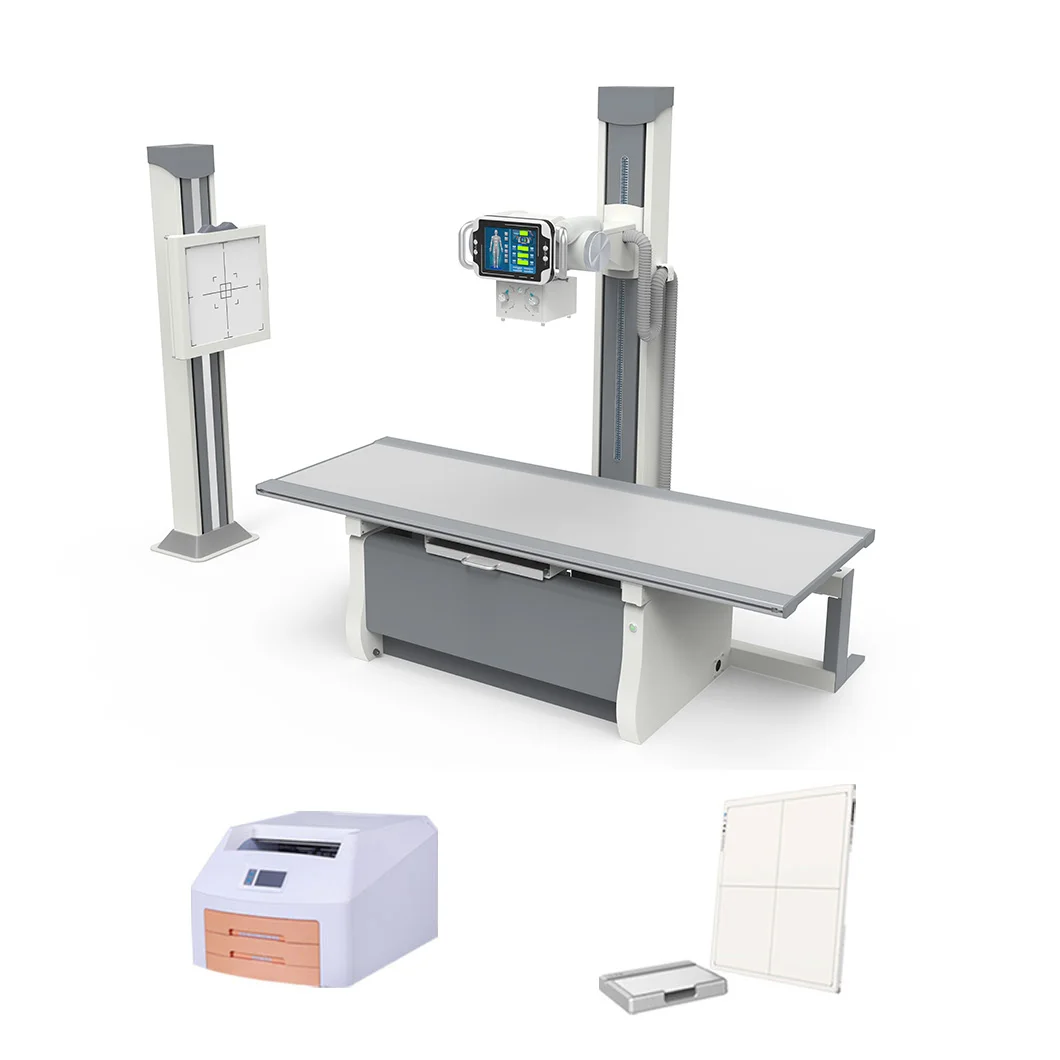
At the heart of an xray machine is a component called the xray tube, which produces the high-energy radiation needed for imaging. This tube contains two main parts: a cathode (negative electrode) and an anode (positive electrode), sealed in a vacuum to prevent energy loss. When the machine is turned on, an electric current heats the cathode, causing it to emit a stream of electrons. These electrons accelerate at high speed toward the anode—usually a tungsten target—due to a strong voltage difference between the two electrodes. When the electrons collide with the tungsten target, their kinetic energy is converted into two forms: heat (most of it) and xray photons (the useful radiation). The xray tube is designed to focus these photons into a narrow beam, which is then directed toward the patient’s body. This controlled generation of xrays is the first critical step in creating diagnostic images.
Xray Beam Penetration and Tissue Interaction
Once the xray beam is generated, it travels through the patient’s body, and this is where the image begins to take shape. Different body tissues absorb xrays at different rates, based on their density and composition. Dense tissues like bones and teeth absorb most of the xray photons, allowing very few to pass through. Less dense tissues such as muscles, fat, and organs absorb fewer photons, letting more pass through. Air-filled spaces like the lungs allow almost all xrays to penetrate. This difference in penetration creates a “shadow” pattern: areas where few xrays pass through (dense tissues) appear light on the final image, while areas where many xrays pass through (less dense tissues) appear dark. For example, a broken bone will show up as a bright white area against the darker background of surrounding muscles and soft tissues. This contrast is what lets doctors distinguish between normal and abnormal structures in the body.
Capturing the Xray Image: Detectors and Screens
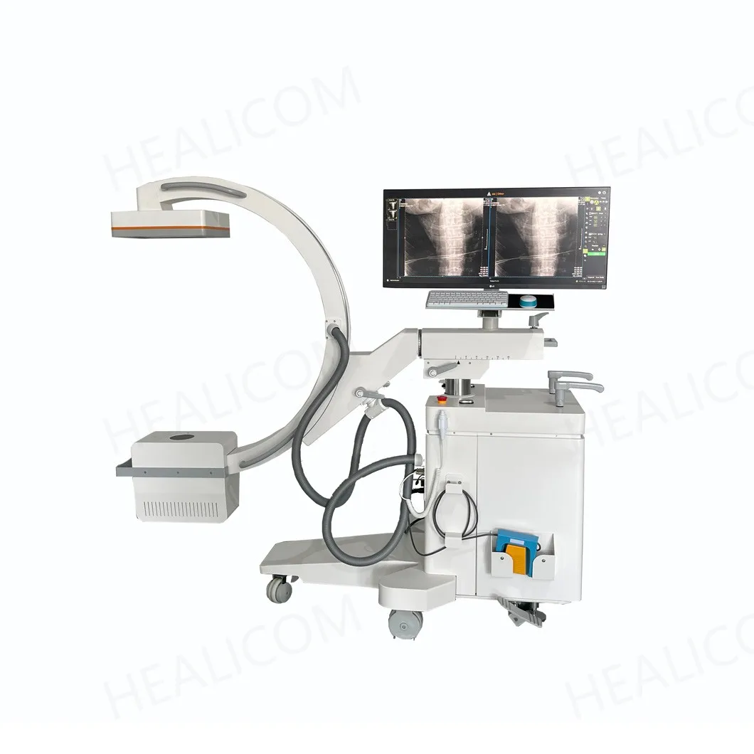
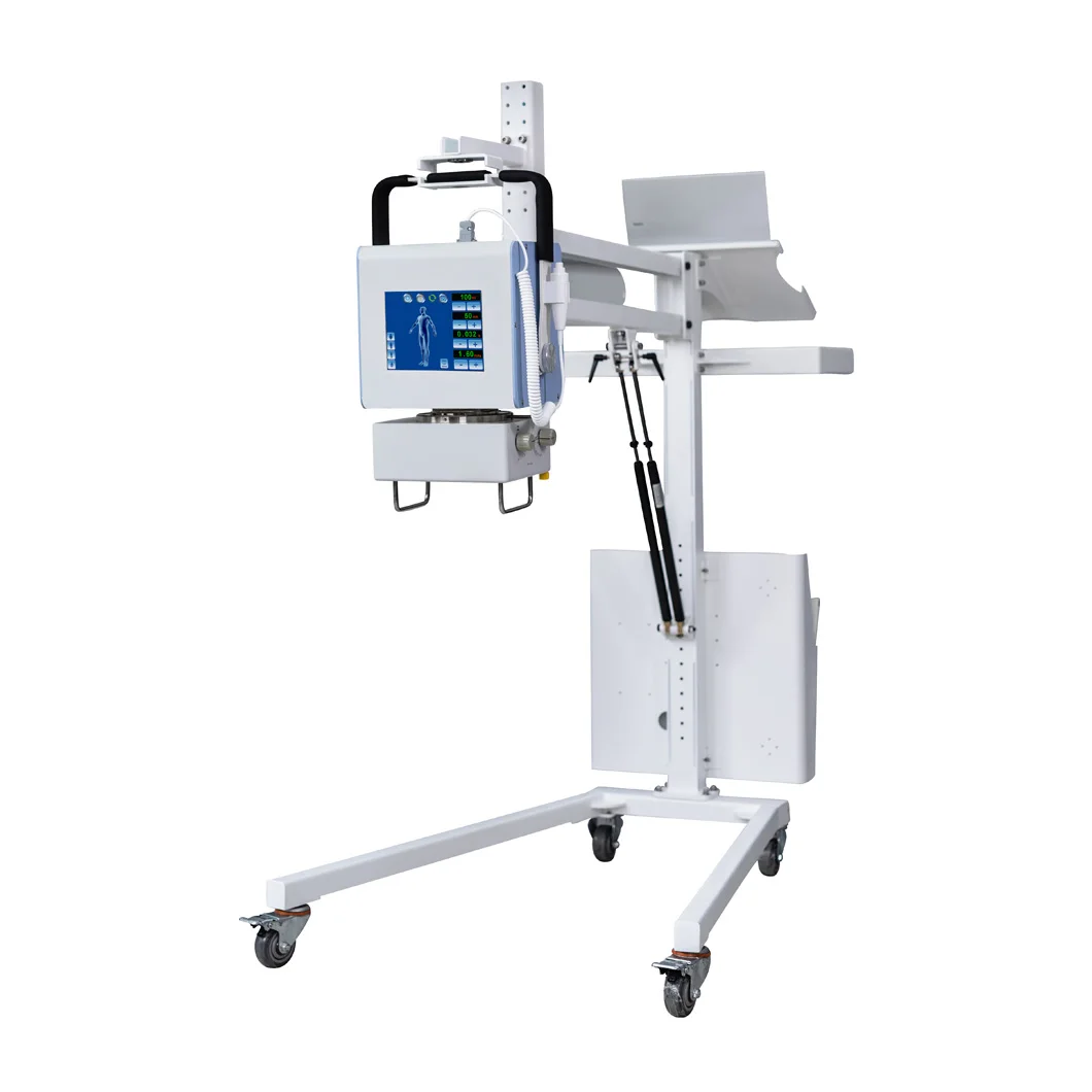
After passing through the patient, the xray beam (now carrying the tissue contrast information) hits an image detector—a key component that converts the radiation into a visible image. Traditional xray machines used film screens: the xrays expose a special film coated with light-sensitive chemicals, which is then developed in a darkroom to reveal the image. Modern digital xray machines, however, use digital detectors that are faster and more efficient. These detectors contain sensors that convert xray photons into electrical signals. The signals are then sent to a computer, which processes them into a digital image displayed on a monitor. Some digital detectors use flat-panel technology, which provides high-resolution images with minimal radiation exposure. Unlike film, digital images can be adjusted—brightened, darkened, or zoomed in—immediately, helping doctors get a clearer view of specific areas. This capture step is crucial for turning the invisible xray beam into a usable diagnostic tool.
Image Processing and Enhancement for Diagnosis
Once the digital image is captured, the xray machine’s computer system performs processing and enhancement to improve its diagnostic value. Raw images may be too dark, too bright, or lack sufficient contrast, so the computer adjusts these parameters to highlight important details. For example, in a chest xray, the software can enhance the contrast between the lungs and the heart to make it easier to spot signs of pneumonia or fluid buildup. Advanced processing techniques can also reduce noise (unwanted graininess) and sharpen edges, making small abnormalities more visible. Digital images can also be analyzed using specialized software—for instance, measuring the size of a tumor or the density of a bone fracture. Additionally, these images can be stored electronically in a hospital’s database, shared with other doctors for second opinions, or printed for patient records. This processing step ensures that the final image is clear, detailed, and tailored to the doctor’s diagnostic needs.
Safety Features and Radiation Control
While generating xrays is essential for imaging, an xray machine also includes built-in safety features to protect both patients and operators from excessive radiation exposure. The machine allows doctors to adjust the dose of radiation based on the patient’s size, age, and the area being imaged—children and small adults receive lower doses than larger adults. Lead shielding, such as aprons and collars, is used to cover parts of the body not being imaged, reducing unnecessary exposure. The xray beam is also collimated (focused) to a specific area, minimizing radiation to surrounding tissues. Modern xray machines are designed to emit radiation only during the actual exposure—usually a fraction of a second—further reducing risk. Operators stand behind lead barriers or use remote controls to operate the machine from a safe distance. These safety measures ensure that the benefits of xray imaging far outweigh the minimal radiation risk, making the xray machine a safe and reliable diagnostic tool.
In conclusion, an xray machine generates diagnostic images through a coordinated process: generating xray radiation via an xray tube, using tissue density differences to create contrast, capturing the beam with digital detectors, enhancing the image through computer processing, and ensuring safety with radiation control features. This combination of physics, technology, and engineering has made the xray machine an indispensable tool in modern medicine. Whether it’s diagnosing a simple fracture or detecting a life-threatening condition, the xray machine’s ability to see inside the body quickly and non-invasively has saved countless lives. As technology advances, xray machines continue to become more efficient, safer, and more precise, further improving their value in medical diagnosis and patient care.

 Hot News