EN


How Can an OCT System Fit Into a Modern Eye Clinic?
An OCT system earns its place in a modern eye clinic when it supports a defined clinical workflow rather than becoming a stand-alone technology purchase. Optical coherence tomography is a non-invasive imaging method that uses light to produce cross-sectional retinal images. For a clinic, that technical capability matters only when the right patient can be scanned at the right point in the visit, the image reaches the appropriate reviewer, and the result can be documented and acted on within local clinical protocols.
That is why an OCT scanner machine should be evaluated as part of a service pathway. More accurately, the question is not simply whether an OCT has an appealing feature list. The question is whether the proposed workflow, staffing, room layout, data route, training plan, and service arrangements make an OCT eye scan reliable in everyday use.
Start with the clinic workflow, not the specification sheet
A useful starting point is to map a typical patient journey: referral or check-in, preliminary testing, dilation where locally appropriate, image capture, clinician review, discussion, documentation, and follow-up. The value of an OCT is linked to how smoothly it fits between those steps. An image taken too late, stored in an isolated location, or unavailable to the reviewing clinician can add effort without improving the clinic’s process.
Optical coherence tomography can allow clinicians to view retinal layers and the optic nerve fiber layer. That helps explain why image quality, repeatability, review access, and local interpretation procedures deserve attention. It does not, by itself, establish a diagnosis or replace professional judgement. A clinic should define which team member captures the scan, who checks image adequacy, who reviews it, and what happens if a patient cannot be scanned successfully on the first attempt.
- List the visit types in which an OCT may be considered.
- Identify the handoff between the technician, optometrist, ophthalmologist, and administrative team.
- Decide whether the OCT eye scan is performed before, during, or after the clinician encounter for each pathway.
- Set local escalation and re-scan procedures rather than assuming every image is usable.
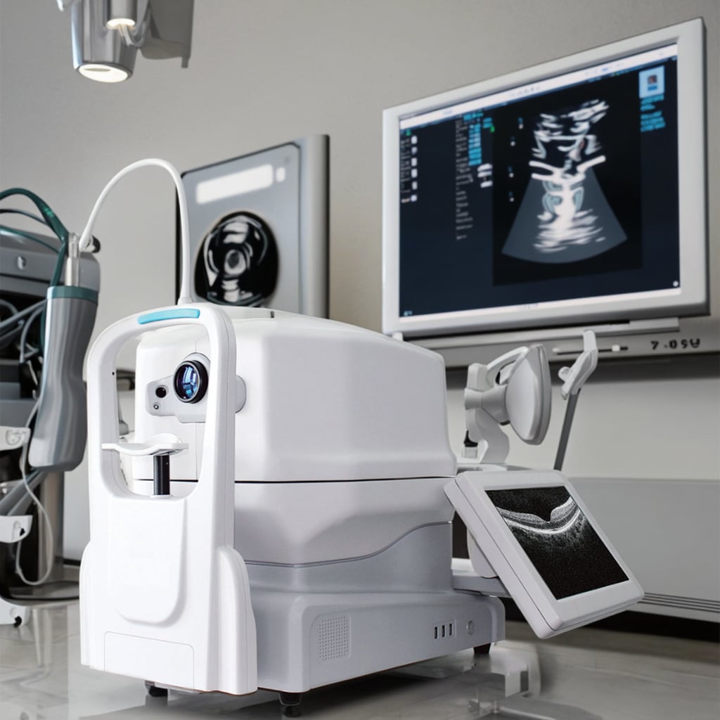 A workflow review can help a clinic define how image capture, review, and documentation fit together.
Choose placement and patient flow deliberately
Where the OCT scanner machine sits changes how it is used. A unit located near preliminary testing may support a structured testing sequence; a unit in a consulting area may suit a clinician-led discussion. Neither arrangement is universally better. The right choice depends on room availability, privacy, wheelchair access, patient movement, and the number of people sharing the equipment.
Before selecting ophthalmic imaging equipment, run a short walk-through with the people who will use it. Check the approach to the device, seating, lighting, cable management, cleaning responsibilities, power provision, network access, and what happens when a room is occupied. A paper workflow can reveal small barriers that a product demonstration may not show. The same exercise should identify where staff will explain the scan to patients and where images will be reviewed without interrupting the next appointment.
Define image, data, and review requirements early
An OCT system should be assessed against the clinic’s actual information flow. Ask how images are identified, stored, retrieved, compared over time, and associated with the patient record. Confirm the required interfaces, user permissions, export options, security controls, retention approach, and responsibilities with the clinic’s IT and governance teams. Compatibility should be verified for the specific proposed configuration rather than inferred from a general marketing statement.
The practical review question is equally important: who needs to see the OCT image, on which screen, and at what stage of the visit? For example, a team may want a technician to complete acquisition while a clinician reviews the study later in the same appointment. Another clinic may need review during a specialty consultation. Map those choices before comparing software options, because a feature only adds value when it supports the workflow step that needs it.
Build training and quality checks into deployment
Adding an OCT is a change-management task as well as an equipment project. Create role-specific training that covers patient positioning, acquisition steps, image-quality checks, hygiene procedures, troubleshooting boundaries, data handling, and escalation. A short competency check after initial training can be more useful than treating attendance as evidence of readiness.
For an OCT eye scan, document the clinic’s local expectations for repeat scans, artefacts, incomplete studies, and clinician review. Include front-desk and scheduling colleagues when their actions affect visit timing. Then schedule a controlled go-live period: observe the first workflows, collect staff questions, and adjust rooming or appointment templates when needed. This approach respects the fact that ophthalmic imaging equipment works within a team, not in isolation.
Evaluate the full ownership plan
Price is one input, but an OCT scanner machine should be assessed across deployment, training, maintenance, software, accessories, service response, and future workflow needs. Ask suppliers to state what is included in the proposed configuration and which items require separate agreement. The clinic should also clarify acceptance criteria before delivery: expected installation steps, the training scope, the tests to be witnessed, documentation to be supplied, and the contact route for service issues.
A structured comparison table can prevent mismatched quotations. Use the same headings for every option: intended clinical tasks, capture workflow, image review, data route, footprint, training, maintenance, service, and acceptance evidence. This helps procurement, clinical, engineering, and management stakeholders discuss the same OCT project without reducing the decision to a single specification.
A practical rollout checklist
- Describe the patient pathways where an OCT is expected to contribute.
- Assign capture, quality-check, review, documentation, and escalation responsibilities.
- Validate room layout, accessibility, power, network, and cleaning arrangements on site.
- Confirm the proposed OCT scanner machine configuration, interfaces, and local data-governance requirements in writing.
- Plan role-based training and a supervised go-live period.
- Agree on acceptance tests, maintenance ownership, and a review date after implementation.
Make the OCT system part of a repeatable service
A modern clinic benefits most when an OCT becomes a dependable part of a defined service, not a separate destination for images. Optical coherence tomography can provide clinically useful visual information, but equipment selection and deployment should remain conditional on the clinic’s patient mix, professional protocols, staffing model, local requirements, and confirmed technical fit. By defining those conditions first, a clinic can evaluate an OCT system with a clearer view of what must work on day one and what should be monitored over time.

 Hot News