EN


What Quality Assurance Checks Are Important for CBCT Equipment?
For a dental practice, clinic, or imaging center, a CBCT quality-assurance program is not a one-time commissioning task. It provides routine evidence that the CBCT system still produces images suitable for the clinical questions it must answer. As a medical-equipment selection adviser, I recommend starting with workflow: decide who performs each CBCT check, what baseline is used, where results are recorded, and what happens when a result changes.
CBCT supports three-dimensional assessment of teeth, jaws, facial bone, and adjacent structures. That usefulness depends on consistent geometry, image quality, and radiation-safety performance. The exact test list, limits, and frequency should follow applicable regulations, the manufacturer’s instructions, and advice from a qualified medical physicist. Still, the categories below give procurement teams and operators a practical way to structure a CBCT QA program.
Start with acceptance testing and a usable baseline
Before routine CBCT use, document acceptance measurements for each clinical protocol and field of view. Acceptance testing establishes the reference against which later constancy tests are compared. Record the CBCT model, software version, detector configuration, reconstruction settings, exposure protocols, phantom, and measurement setup. If a firmware update, detector repair, tube replacement, or relocation changes the system, repeat the relevant baseline checks.
This matters because a CBCT image can look acceptable at a glance while a gradual change in noise, uniformity, or geometry has already affected a small-field protocol. A stable baseline lets the team identify a trend before it becomes a service interruption. A qualified physicist can help define control limits that make sense for the particular CBCT unit rather than borrowing values from a different platform.
1. Check operation, safety, and mechanical condition
Daily or before-use CBCT checks are usually simple. Confirm that warning lights, exposure indicators, emergency stops, interlocks, patient supports, positioning lasers, displays, and controls operate as intended. Inspect cables, detector covers, head supports, and moving components for damage or unusual motion. Verify that the CBCT warm-up and self-test complete without unexpected messages.
Positioning deserves attention because a CBCT scan is only as reliable as its relationship to the intended anatomy. Check that localization lights and patient-support references align with the documented setup. A mismatch can lead to repeated scans, an unsuitable field of view, or a study that does not answer the referral question. Log faults even if they clear after restarting; intermittent CBCT behavior is useful information for service personnel.
2. Monitor image uniformity, gray values, and noise
A homogeneous phantom is central to CBCT constancy testing. With the same phantom placement and acquisition protocol, review uniformity across the reconstructed volume, mean pixel or gray-value behavior where relevant, and image noise. These checks look for shading, rings, streaks, nonuniform regions, or a drift from the established CBCT baseline.
Uniformity is not merely an engineering measure. In maxillofacial imaging, nonuniformity can make subtle density differences harder to interpret or can obscure whether a change comes from anatomy or the acquisition chain. Noise should also be trended, rather than judged from a single image. A sudden rise may point to detector, calibration, exposure, reconstruction, or environmental issues. Use the phantom and analysis method specified for that CBCT system whenever possible.
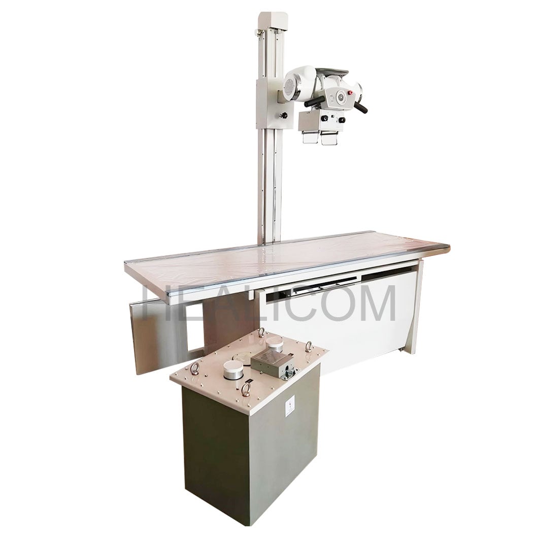 A CBCT QA workflow uses repeatable phantom imaging and documented image review.
3. Verify contrast, spatial resolution, and voxel-size performance
Image-quality checks should match the tasks for which the CBCT equipment is used. Low-contrast checks assess whether modest differences in attenuation remain distinguishable under defined conditions. High-contrast spatial-resolution checks assess whether fine structures can be separated. Test patterns, line-pair inserts, or other manufacturer-approved phantom features can provide repeatable evidence.
Voxel size must be interpreted carefully. A smaller voxel size can support finer sampling, but it does not independently establish clinical detail; motion, noise, reconstruction, field of view, and exposure selection also matter. For CBCT protocols used in endodontic, implant, orthodontic, or broader maxillofacial imaging workflows, confirm that the displayed voxel size, reconstruction setting, and phantom result remain consistent with the baseline. Do not assume that selecting the smallest voxel size is appropriate for every referral.
4. Look for artifacts and geometric accuracy
Routine CBCT review should include artifacts that can affect interpretation: rings, streaks, banding, truncation, motion-related blur, and reconstruction anomalies. Compare a standard phantom image with the baseline image under the same protocol. If the artifact pattern changes, preserve the images and document the protocol, operator, and service status before changing multiple variables at once.
Geometric accuracy is equally important. Check dimensions or known phantom distances using the CBCT viewer and its documented measurement procedure. Confirm localization-light accuracy and, where applicable, alignment between scout/localizer information and the planned volume. These checks help ensure that a CBCT system is not quietly shifting the relationship between displayed anatomy and physical space.
5. Review exposure settings and radiation-output oversight
Exposure and dose-related checks should be performed by appropriately qualified personnel using suitable instruments and local requirements. The aim is to confirm that selected CBCT protocols, collimation, field of view, and displayed exposure information remain consistent with the approved baseline. Verify that protocols are named clearly enough for operators to select the intended clinical task and patient category.
Do not substitute a generic dose target for a CBCT-specific review. The appropriate approach depends on equipment design, protocol, field size, regulation, and clinical purpose. When a CBCT protocol is added or modified, involve the responsible physicist and document the change-control decision. This supports optimization without claiming that one setting fits every dental imaging equipment workflow.
6. Use a schedule, trend records, and action limits
A useful CBCT QA plan separates daily functional checks, periodic phantom constancy checks, annual or physicist-led evaluations, and event-triggered checks after repair or software change. The New Jersey dental CBCT QA manual, for example, illustrates daily functional, water/gray-value, and uniformity checks, with initial and annual image-quality checks; it should not be treated as a universal legal schedule. Your local program may require different frequencies.
| QA category | Typical evidence | When to escalate |
|---|---|---|
| Functional and safety | Indicators, interlocks, supports, positioning | Failure, intermittent behavior, or unusual motion |
| Phantom image quality | Uniformity, noise, contrast, resolution, artifacts | Drift beyond local action limits or new artifact patterns |
| Geometry and setup | Known distances and localization accuracy | Mismatch from baseline or repeat-positioning issues |
| Protocol governance | Approved settings, change log, training records | New protocol, software update, repair, or unexplained outcome |
Keep CBCT records in a form that allows trends to be seen: date, operator, protocol, phantom, measured result, pass/fail decision, corrective action, and follow-up result. A pass/fail box alone is less useful than a trend chart or table that shows slow deterioration.
What to ask a CBCT machine supplier before purchase
A CBCT machine supplier should be able to explain the acceptance process, routine QA instructions, recommended phantom, service response route, software-update process, operator training, and documentation supplied with the system. Ask which checks the operator can perform, which require a physicist, and how baseline data are exported or retained. Request clear protocol documentation for the clinical applications your team expects to perform.
For dental imaging equipment procurement, compare the support model as carefully as the specification sheet. A CBCT unit with a documented QA pathway, accessible service records, and training aligned to your maxillofacial imaging workflow is easier to manage over its operating life. The objective is not to make a CBCT program burdensome; it is to make deviations visible, traceable, and actionable.
Conclusion
Important CBCT quality-assurance checks cover safe operation, positioning, image uniformity, gray-value stability, noise, contrast, spatial resolution, voxel-size consistency, artifacts, geometry, and exposure oversight. Establish a baseline at acceptance, assign responsibilities, trend phantom results, and repeat relevant tests after material changes. By connecting CBCT checks to the clinical protocols actually in use, a practice can maintain a more dependable imaging process while following its local regulatory and professional requirements.
 Hot News