EN


Which Specifications Matter When Comparing OCT Machines?
When an eye clinic compares an OCT machine, a long specification sheet can obscure the real decision. Optical coherence tomography uses light waves to produce cross-sectional retinal images, but a useful OCT purchase decision depends on more than the highest headline number. The right system is the one whose scan protocols, image quality, reporting, and support requirements fit the clinic’s daily work.
For a hospital procurement team, it helps to split the OCT decision into three stages: define the clinical need, compare configurations on the same basis, and verify performance before acceptance. This approach gives clinicians, technicians, IT staff, and purchasers a common evaluation sheet instead of separate assumptions.
Start with the clinical and workflow question
Before comparing an OCT specification, identify the scans the clinic expects to perform, the patient populations involved, and who will review the output. Retina, glaucoma, cornea, and anterior-segment workflows may prioritize different scan patterns, fields of view, segmentation tools, and report layouts. An OCT feature matters only if it supports a defined protocol and a team member can use the result consistently.
Ask for a demonstration using representative scenarios, including difficult fixation, media opacity, or repeat follow-up scans where appropriate. This is also where eye clinic imaging requirements become concrete: room layout, patient positioning, operator time, data storage, and the handoff of reports to the clinical record all affect whether an OCT system fits the day.
Compare resolution and scan speed as a pair
Axial resolution describes the system’s ability to distinguish structures along the depth axis. Scan speed, often expressed as A-scans per second, influences how quickly an OCT system captures data and can affect motion sensitivity, volume coverage, or averaging options. Neither value should be evaluated alone.
Published technology comparisons commonly place spectral-domain OCT examples around 5–7 µm axial resolution and 20,000–52,000 A-scans per second, while swept-source examples may operate at higher scan speeds. These are technology-class reference points, not a universal purchasing threshold. Wavelength, scan protocol, field size, averaging, software processing, fixation, and the stated measurement method can change the practical image result.
When reviewing OCT scan specifications, ask vendors to show the same scan type, scan area, and averaging setting for each candidate system. A faster number is meaningful only when the requested protocol still produces repeatable images that clinicians find usable.
Look beyond the headline scan
| Specification area | What to compare | Verification question |
|---|---|---|
| Scan coverage | Available line, raster, volume, radial, and wide-field protocols | Which protocols match the clinic’s documented use cases? |
| Image quality | Signal quality indicators, artifact handling, repeatability, and averaging behavior | Can the team review representative images under the same protocol? |
| Segmentation and analysis | Layers, maps, trend tools, edit controls, and error flags | How are segmentation errors found, corrected, and recorded? |
| Reports and data | Report formats, export options, archive capacity, and workflow integration | Who receives the report, and can it be retrieved at follow-up? |
| Serviceability | Training, maintenance expectations, software updates, and response process | What is documented before installation and at acceptance? |
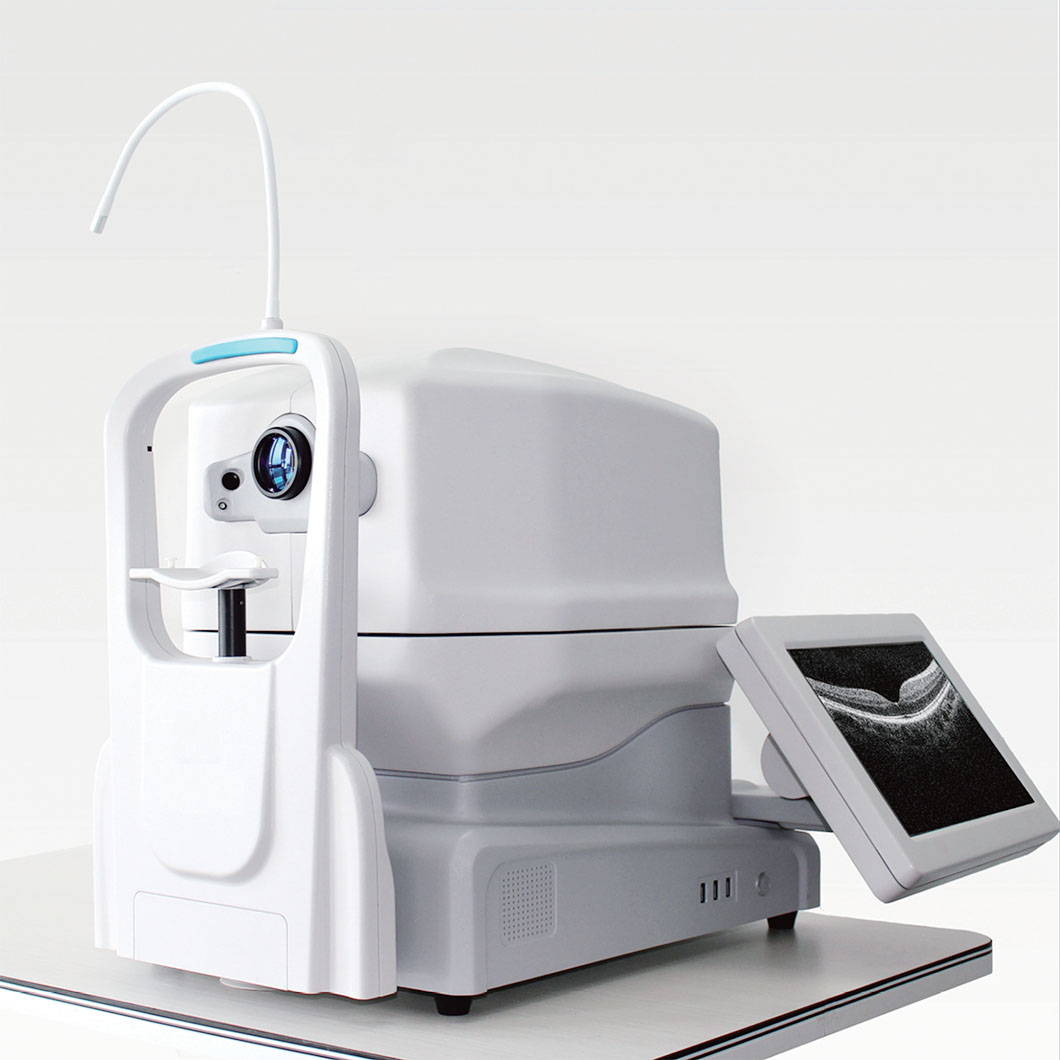 An abstract retinal-layer visual supports a like-for-like review of OCT scan protocols.
Check scan coverage, optics, and patient access
A candidate OCT system may offer several scan sizes or specialties, yet the relevant comparison is the coverage required by the service line. If a clinic needs macular volume scans, optic-nerve analysis, or anterior imaging, request the exact protocols rather than relying on a general modality label. For an OCT equipment supplier, a clear requirement list reduces the risk of comparing optional modules with a base configuration.
Also consider working distance, chin-rest adjustment, fixation targets, alignment aids, and whether the system supports the clinic’s expected patient mobility needs. A broadly configurable OCT platform can cover more scenarios, but a specific patient group or room workflow may need additional confirmation. Record what was demonstrated and what still requires validation.
Evaluate software as carefully as hardware
In routine ophthalmic diagnostics, clinicians often depend on analysis displays, longitudinal comparison tools, and report consistency as much as on raw image acquisition. Review the user permissions, segmentation correction workflow, language options, export format, and data-retention approach. Your team should also clarify whether software updates could alter measurements, reports, or workflow and how those changes are communicated.
Change the question from “Does this OCT have analysis software?” to “Can our users locate, review, correct, and communicate the information needed for this protocol?” That framing avoids treating a feature list as proof of workflow fit.
Make image quality and acceptance testing visible
Image quality should be demonstrated rather than assumed from resolution alone. The FDA has described a retina phantom with model eye as a regulatory science tool for ophthalmic OCT image-quality assessment, which is a useful reminder that repeatable evaluation matters. It does not establish that a particular device is cleared for a particular use or replace the clinic’s own acceptance criteria.
Before installation, agree on a practical acceptance checklist: delivered configuration, selected scan protocols, image display, report generation, user training, connectivity testing, and maintenance contacts. If preventative maintenance responsibilities and software support are not discussed early, accountability can become unclear after deployment.
Use a like-for-like procurement matrix
Place each OCT option in the same matrix: configured hardware, available protocols, quoted software modules, implementation scope, training, maintenance, and evidence from the demonstration. Include initial cost alongside deployment, training, maintenance, and recurring resource questions, without assuming a clinic’s budget or assigning a cost outcome.
The final choice should connect each OCT specification to a clinical task, a responsible user, and an acceptance check. For eye clinic imaging, that traceability is more useful than an isolated claim about speed or resolution. It gives the clinic a defensible basis for requesting comparable proposals and for planning a structured evaluation with its preferred OCT equipment supplier.
Conclusion
The specifications that matter most when comparing an OCT machine are the ones that can be tied to a real protocol: appropriate scan coverage, verified image quality, usable analysis software, reliable reporting, patient access, and a documented support plan. Review OCT scan specifications in the same configuration and ask each supplier to demonstrate the workflow your clinic intends to run. That turns an OCT comparison from a checklist of numbers into a controlled procurement decision.

 Hot News