EN


Digital X-Ray vs Traditional X-Ray: What Are the Differences?
Choosing an x ray machine is no longer only a question of generator power or room layout. For a hospital, clinic, or imaging center, the real decision is how the image is captured, reviewed, stored, and controlled within the daily workflow. Digital and traditional film-based radiography use X-rays to create projection images, but their image receptors and downstream processes are very different.
From a procurement perspective, compare each x ray machine against the examinations your team performs, the available infrastructure, staffing, image-review needs, and quality-assurance plan. A digital system is often evaluated for speed and connectivity, while a film-based setup may be considered where an existing process still relies on film. Neither label alone settles the purchasing decision.
The core difference: how an X-ray image is captured
A traditional radiography workflow records the exposure on film. The film is then chemically processed, dried, checked, and physically filed or digitized for later sharing. This creates a tangible record, but it also adds handling steps and depends on consistent processing conditions.
A digital X-ray system uses an electronic image receptor. In computed radiography, a cassette with a photostimulable plate is taken to a reader after the exposure. In direct digital radiography, a radiography detector sends image data to a workstation without the film-development stage. The image can then be reviewed, adjusted within permitted processing controls, and routed through the facility’s information workflow.
The distinction matters because the detector is only one part of the x ray machine workflow. Generator settings, positioning, exposure technique, display calibration, network availability, archive rules, and staff training all influence whether a new system fits the department.
Digital vs traditional X-ray at a glance
| Decision area | Digital radiography | Film-based radiography |
|---|---|---|
| Image receptor | Electronic detector or reusable plate | Radiographic film and processing materials |
| Image availability | Available after acquisition and system processing | Available after film processing and handling |
| Review and sharing | Supports workstation review and electronic routing when local systems are configured | Relies on physical film handling unless images are later digitized |
| Consumables | May reduce film and chemical use, but requires detector care and service planning | Requires film, chemicals, processing equipment, and storage materials |
| Quality controls | Requires detector, display, exposure-indicator, and software workflow checks | Requires film, processor, chemical, and viewing-condition checks |
This table is a starting point rather than a substitute for local technical evaluation. A x ray machine should be compared with like-for-like scope: detector size, generator configuration, mobile or fixed use, workstation software, service coverage, and required integration should be defined before quotations are compared.
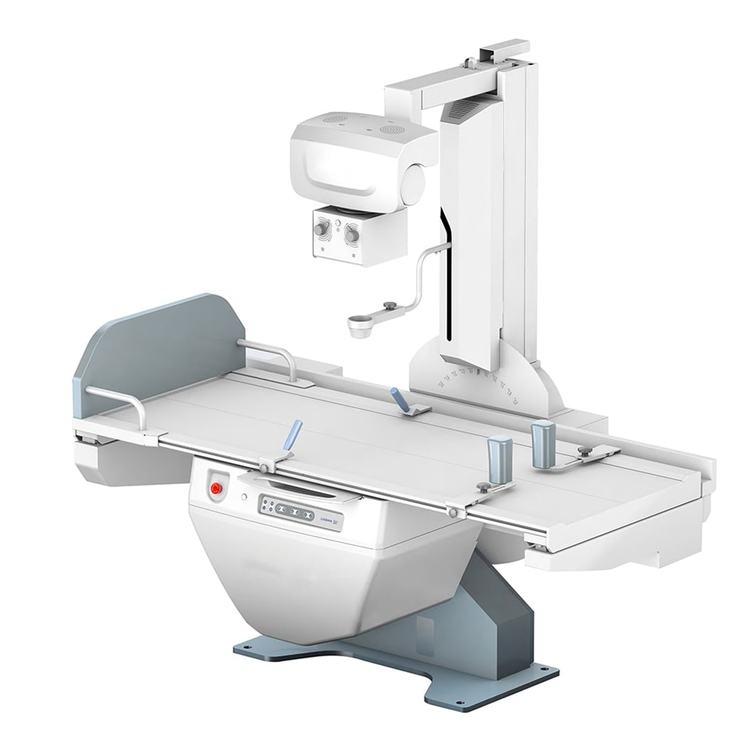
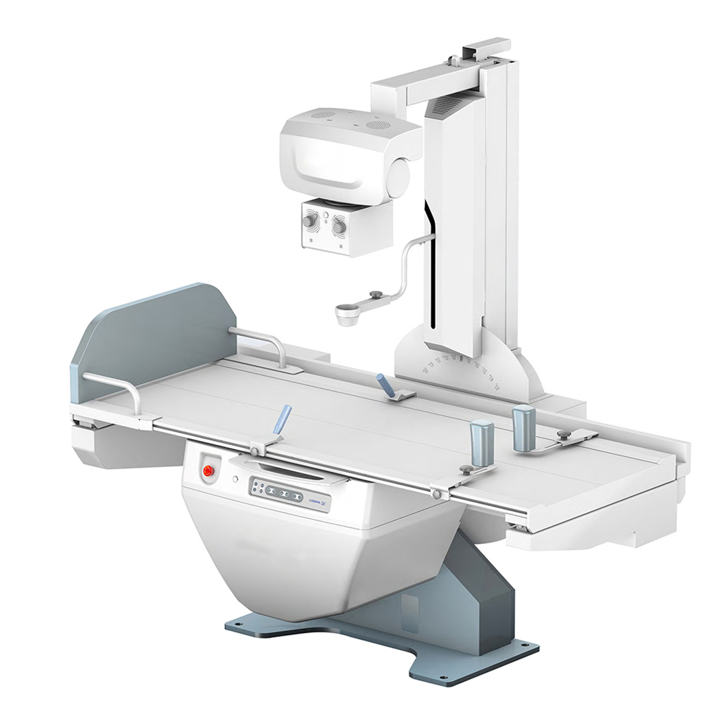 Digital image capture adds detector and workstation checks to the radiography workflow.
What changes in the clinical and operational workflow?
With digital radiography, staff can review image availability soon after acquisition and can route images electronically when the facility has compatible systems and permissions. That can simplify communication between acquisition, review, and archive stages. It also changes the training need: operators must understand detector handling, workstation functions, reject analysis, and the department’s protocol for image labeling and transmission.
Film-based radiography has a different set of dependencies. Film selection, darkroom or processor conditions, chemical management, artifacts, and physical storage become routine controls. Some sites may retain film workflows for established operational reasons, but teams should account for the ongoing supplies, processing environment, and retrieval process—not only the initial equipment cost.
A portable X-ray machine adds another layer to either workflow. Mobility can support bedside examinations, but the buyer should examine battery or power arrangements, detector protection during transport, infection-control procedures, wireless reliability where applicable, and how images are identified and transferred outside the main room. Portability does not remove the need for defined operating procedures.
Image quality, dose, and radiation protection
Both technologies use ionizing radiation for projection radiography. The U.S. FDA describes justification and optimization as radiation-protection principles for medical X-ray imaging. In practical terms, the examination should be clinically justified, and technique, positioning, collimation, shielding practices where locally indicated, and repeat-exam controls should be managed by qualified professionals under applicable requirements.
A common purchasing mistake is to treat a wider digital exposure latitude as permission to be less disciplined about technique. It is not. A digital detector may produce an image that appears usable across a range of exposures, making exposure monitoring and reject review important. The IAEA’s quality-assurance material connects appropriate image quality with dose monitoring and reduction. Your x ray machine evaluation should therefore ask how the system displays exposure information, supports audit records, and fits the facility’s quality program.
Radiation protection is not a marketing feature attached to one modality. It is a coordinated responsibility involving equipment performance, room design, protocols, training, maintenance, and oversight. Local regulations and the facility’s radiation-safety professionals should determine the controls required for a particular installation and examination mix.
How to compare costs beyond the purchase price
Digital systems may shift spending from film and chemistry toward detectors, workstations, networking, software, service, and cybersecurity or archive planning. Film systems may have a lower apparent transition burden where processing already exists, yet supplies, processor upkeep, environmental handling, and physical records can remain recurring costs. The useful comparison is a lifecycle estimate, not a single equipment line item.
For each x ray machine proposal, ask suppliers to separate included and optional items: generator, stand or table, detector count and size, workstation, acquisition software, installation scope, training, acceptance testing support, warranty, response terms, and planned maintenance. This avoids comparing a complete digital X-ray system with a base configuration that omits essential workflow components.
A practical selection checklist
- List the examination types, patient locations, throughput patterns, and image-routing needs.
- Define whether a fixed room, a portable X-ray machine, or both are needed.
- Specify detector dimensions, durability expectations, backup arrangements, and handling procedures.
- Review the planned quality-assurance schedule for the generator, radiography detector, displays, software, and repeat analysis.
- Confirm site readiness, power, room shielding evaluation, network access, and acceptance responsibilities with qualified local teams.
- Compare service documentation, training scope, parts availability, and escalation paths alongside the quoted configuration.
Which option should you choose?
A digital x ray machine may fit organizations that need electronic image handling, faster image availability, and an integrated review workflow, provided they can support the associated detector, IT, and QA requirements. Traditional film radiography may remain relevant where an established film workflow and local support structure are the governing constraints. The more accurate conclusion is not that one approach is universally superior; it is that the chosen system must match the examinations, operating environment, and governance capacity of the facility.
Before finalizing a purchase, turn this comparison into an acceptance checklist. Confirm the exact configuration, test it against representative workflow scenarios, and have the appropriate clinical, engineering, radiation-safety, and procurement stakeholders review the plan. That process gives an x ray machine decision a firmer basis than a specification sheet alone.


 Hot News