EN


How Does OCT Imaging Support Retinal Examination?
In a retinal examination, OCT gives the clinician a cross-sectional view that is difficult to obtain from a surface view alone. Optical coherence tomography uses reflected light to build detailed slices through retinal tissue. It does not replace a dilated examination, patient history, or clinical judgment. Instead, it adds a repeatable structural record that can help an eye-care team ask more precise questions about what they are seeing.
For clinics evaluating ophthalmology diagnostic devices, the value of OCT is not simply that it produces an image. The useful question is whether a retinal OCT scan is captured consistently, interpreted in context, and comparable with prior examinations. That distinction matters when the goal is assessment, monitoring, referral, or documentation rather than a standalone diagnosis.
What OCT adds to a retinal examination
A routine retinal examination lets a clinician assess the back of the eye directly. OCT complements that view by presenting sectional anatomy of the retina. In a normal scan, individual retinal layers can often be distinguished. When a layer’s contour, thickness, or reflectivity appears different from the expected pattern, the image can guide the next clinical step.
This structural perspective can be useful when symptoms, fundus findings, and visual function do not tell the same story. For example, a clinician may use OCT to look for fluid, traction, a change in the foveal contour, or disruption near the retinal pigment epithelium. The scan itself does not name the cause. It creates evidence that must be considered alongside examination findings and, when appropriate, other tests.
How a retinal OCT scan fits into clinical workflow
A practical OCT workflow begins before the image is captured. The operator confirms patient positioning, fixation, and the intended scan location. After acquisition, the reviewer checks that the retinal area of interest is centered and that segmentation lines and scan boundaries make sense. This is where a retinal OCT scan becomes more than a picture: it becomes a usable record for the clinical encounter.
- Acquire: capture the appropriate scan pattern while helping the patient maintain fixation.
- Verify: review signal strength, motion, blink effects, decentration, and segmentation behavior.
- Correlate: compare the OCT appearance with symptoms, visual acuity, examination findings, and prior images.
- Document and follow: retain comparable scans when follow-up is clinically indicated.
That sequence is important because a technically impressive instrument cannot compensate for a poorly centered or artifact-heavy image. A clinician may need to repeat a scan or adjust the acquisition approach before drawing conclusions.
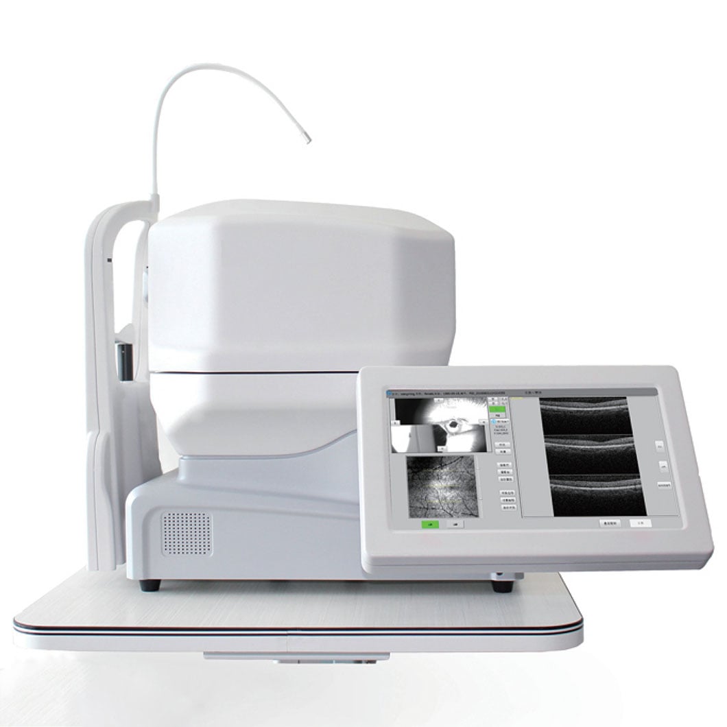 An illustrative OCT imaging scene; clinicians interpret scans in the context of the full examination.
Why OCT image quality affects interpretation
OCT image quality directly affects confidence in what can be seen. Media opacity, dry eye, small pupils, poor fixation, blinking, motion, or an off-center scan can reduce clarity. Automated layer segmentation can also be imperfect, particularly when anatomy is altered. A colored thickness map or numerical value should therefore be checked against the underlying B-scan rather than accepted in isolation.
For procurement teams, this creates a practical evaluation criterion for retinal imaging technology: look beyond headline resolution. Consider how the device displays scan quality, supports repeatable positioning, shows raw B-scans, handles follow-up comparison, and fits into local image-review and recordkeeping procedures. Training needs and service support also influence whether an OCT system can be used reliably day after day.
Structural OCT and OCT angiography are related, not interchangeable
Standard structural OCT describes retinal and choroidal anatomy through cross-sectional imaging. OCT angiography, often called OCTA, uses repeated scans to visualize motion contrast associated with blood flow in selected retinal or choroidal layers. OCTA can add information in suitable clinical situations, but it has its own artifacts and limitations. Neither modality should be treated as an automatic substitute for examination, dye-based testing when indicated, or clinician interpretation.
Keeping that distinction clear prevents a common purchasing and workflow error: treating every OCT capability as if it answered the same clinical question. The preferred scan protocol depends on the patient, the clinical concern, and the practitioner’s established standards.
Questions to ask when assessing ophthalmology diagnostic devices
- Which retinal regions and scan patterns are most relevant to the clinic’s patient population?
- How clearly does the system show acquisition quality, motion artifacts, and segmentation errors?
- Can users compare like-for-like follow-up scans without adding unnecessary workflow steps?
- How are images stored, exported, reviewed, and protected within the clinic’s existing processes?
- What training, installation, maintenance, and local regulatory verification are required before use?
These questions keep evaluation focused on clinical workflow rather than feature lists alone. A higher specification is not automatically a better fit if the equipment cannot be integrated, maintained, or reviewed consistently by the intended team.
What patients and clinics should remember
For patients, an OCT examination is typically noninvasive and quick. The care team may use it to clarify a finding, establish a baseline, or monitor a known retinal condition. The result should be discussed with the qualified clinician who has the full examination context.
For clinics, OCT is most helpful when it supports repeatable imaging, careful review, and meaningful comparison over time. Reliable retinal imaging technology combines capable hardware with good acquisition habits and clinical governance. In that setting, OCT contributes a detailed structural layer to retinal examination without overstating what any single image can prove.
Conclusion
OCT supports retinal examination by converting reflected light into cross-sectional retinal images that clinicians can interpret alongside the rest of the patient assessment. A dependable retinal OCT scan can reveal structural detail, support monitoring, and improve documentation. The quality of the image, the integrity of the workflow, and the clinician’s interpretation remain central. When evaluating ophthalmology diagnostic devices, choose a system whose image quality controls, review tools, and service pathway match the real conditions of use.
 Hot News