EN


What Are the Main Clinical Applications of OCT Scanners?
In ophthalmology, OCT (optical coherence tomography) gives clinicians a non-invasive cross-sectional view of eye structures. It is usefully understood as a high-detail imaging tool that adds structural evidence to the history, examination, visual-function tests, and clinical judgment. For an eye clinic imaging workflow, the useful question is not simply whether a scanner has OCT, but which clinical decisions the OCT images need to support.
From a clinical-workflow perspective, OCT is most valuable when the image answers a repeatable question: Is retinal anatomy changing? Is fluid present or resolving? Is there a structural pattern that warrants closer assessment? Is the optic nerve or anterior segment better documented today than at the prior visit? OCT does not diagnose a patient by itself, and interpretation remains the responsibility of qualified eye-care professionals. Still, its ability to display retinal layers and the optic nerve fiber layer explains why OCT has become central to many ophthalmic diagnostics pathways.
Why OCT fits into clinical eye imaging
OCT uses light to create cross-sectional images rather than relying on a standard color photograph alone. In routine practice, OCT images can make subtle structural changes easier to document, compare over time, and discuss across clinicians. More accurately, the point is not the number of OCT features, but whether those features are needed by the target workflow.
For clinics, the strength of OCT is its role in a sequence: capture an image, review it alongside symptoms and examination findings, compare it with prior scans when appropriate, and use the combined information to guide follow-up or referral. That sequence is relevant in retina services, glaucoma care, and selected anterior-segment assessments. It is also why image quality, repeatability, data review, and staff training matter alongside headline OCT scan specifications.
1. Retina and macular disease assessment
The most familiar clinical application of OCT is posterior-segment imaging, especially the retina and macula. Cross-sectional OCT views can help an ophthalmologist examine retinal layers and identify structural features that may be relevant in conditions affecting central vision. In a patient with suspected or known macular disease, OCT is commonly used as part of the assessment and monitoring process rather than as a stand-alone answer.
For example, the National Eye Institute describes OCT as a way to image the retina and other parts of the back of the eye when assessing macular edema, helping a doctor see the amount of swelling. In practice, this makes OCT useful when the care team needs a consistent structural record before, during, or after management decisions. A clinician may look for changes in retinal contour, fluid-related findings, or other anatomy that should be interpreted in the patient’s full clinical context.
OCT also supports structured follow-up in retinal conditions such as diabetic eye disease and age-related macular degeneration. The value is longitudinal: comparable OCT examinations can make it easier to recognize a meaningful change, while acknowledging that image artifacts, segmentation behavior, and patient cooperation can affect what is seen. An eye clinic imaging protocol should therefore define who captures OCT, who reviews it, and how prior scans are retrieved for comparison.
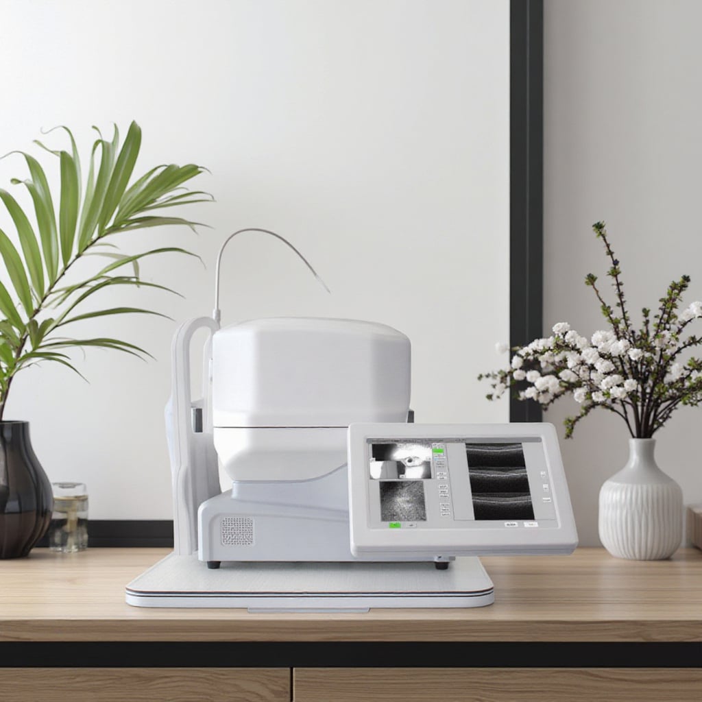 Editorial illustration of OCT supporting retinal image review in an eye clinic.
2. Glaucoma and optic nerve evaluation
Another major application of OCT is assessment related to glaucoma and the optic nerve. OCT can display the optic nerve fiber layer, giving clinicians structural information that may complement pressure measurements, visual-field testing, optic-disc examination, and risk factors. OCT findings must be interpreted carefully: a normal-looking scan does not settle every clinical question, and an abnormal signal can require confirmation against image quality and other evidence.
In a glaucoma workflow, OCT can help establish a baseline and then support trend review. This is particularly helpful when a clinician needs to distinguish a possible structural change from ordinary scan-to-scan variation. Your team should decide who will review the image-quality indicators, whether the same scan protocol will be used at follow-up, and how reports will be incorporated into the medical record. Those operational questions often matter as much as nominal OCT scan specifications.
3. Anterior segment, cornea, and angle imaging
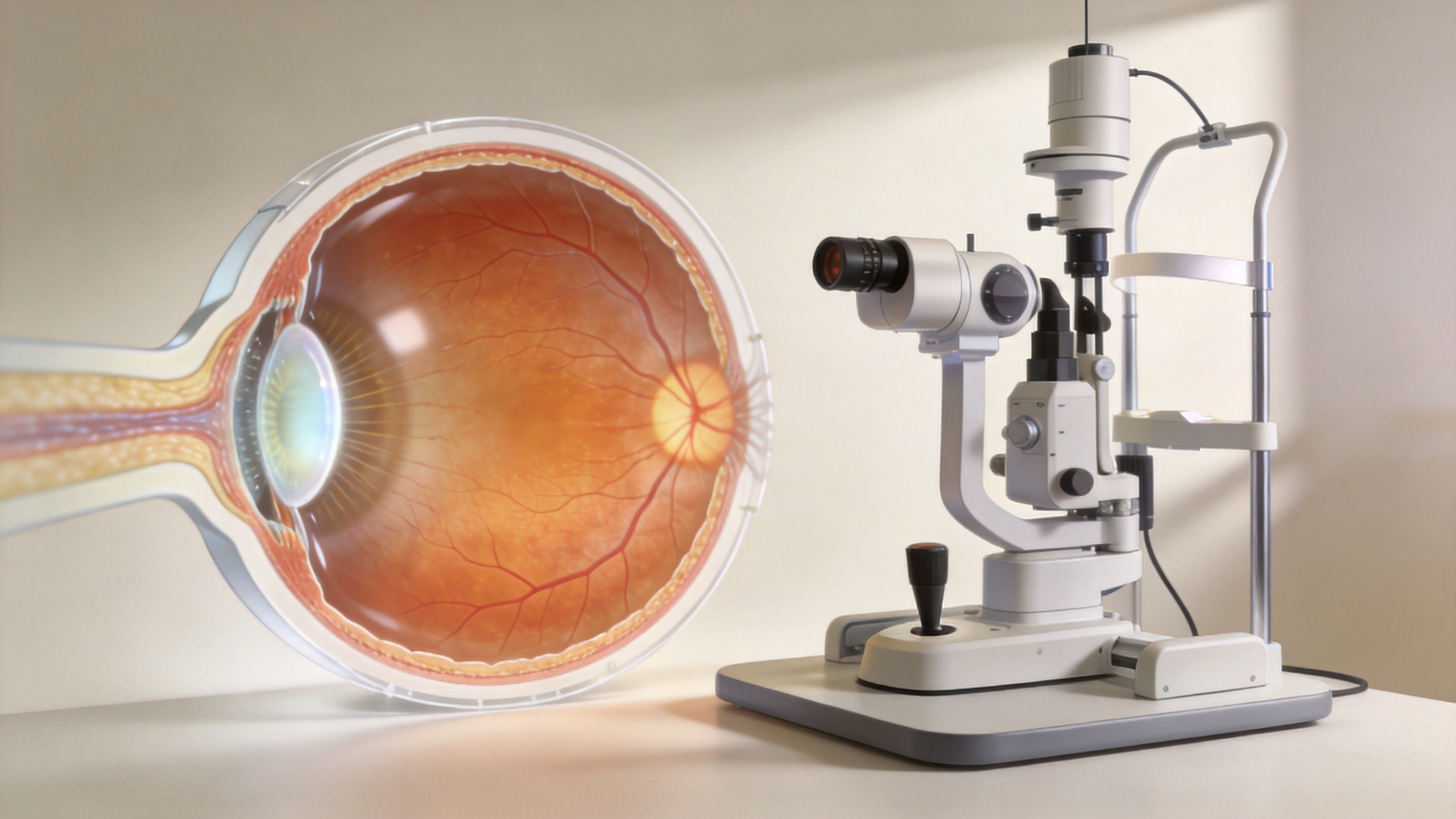
Although many people associate OCT with the retina, anterior-segment OCT has clinical applications for structures including the cornea, conjunctiva, sclera, and anterior chamber. The exact role depends on the device, the scan geometry, the patient, and the clinical question. Anterior-segment OCT may be considered when clinicians need high-detail cross-sectional information about the front of the eye, such as corneal architecture or angle-related anatomy.
This is where a clinic should avoid assuming that all OCT platforms serve the same purpose. A retinal OCT workflow and an anterior-segment OCT workflow can have different requirements for fields of view, scan protocols, documentation, and operator training. If an OCT equipment supplier is discussing a system for anterior-segment use, the clinic should ask for evidence and demonstrations tied to its intended examinations rather than infer performance from a posterior-segment application.
4. Treatment planning and follow-up documentation
Across these clinical areas, OCT contributes most reliably as a documentation and comparison tool. It can help a clinician record anatomy at a point in time, review a change against earlier imaging, and communicate why an assessment or follow-up plan is being considered. In retinal care, OCT may inform the discussion around fluid or layer changes. In glaucoma care, OCT may contribute structural context alongside functional tests. In anterior-segment care, OCT may add cross-sectional anatomy when the clinical question calls for it.
The boundary matters: OCT should not be presented as an autonomous treatment recommender. The interpretation of any OCT result depends on patient symptoms, examination, relevant testing, image quality, and clinician judgment. This discussion is a selection and workflow framework, not a substitute for local regulations, clinical protocols, or professional assessment.
What should an eye clinic compare before adopting OCT?
For procurement teams, clinical applications should lead the comparison. Rather than starting with a feature list, build a short set of workflow questions:
- Clinical scope: Will OCT be used mainly for retina, glaucoma-related assessment, anterior-segment imaging, or a defined combination?
- Image review: Who captures OCT scans, who validates scan quality, and who reviews outputs during a busy clinic session?
- Repeatability: Can the selected protocol be used consistently enough to support meaningful follow-up comparisons?
- Data pathway: How will OCT images, reports, and prior examinations be stored, retrieved, and reviewed within the clinic’s systems?
- Training and support: What onboarding, protocol guidance, maintenance pathway, and local service arrangements will be available?
These questions make eye clinic imaging decisions more practical. They also give an OCT equipment supplier a clearer brief: show the workflow, explain the documented capabilities, and identify any requirements that still need local verification. A supplier discussion is strongest when it connects OCT scan specifications to the clinical tasks the department actually performs.
Key takeaway
The main clinical applications of OCT scanners are retinal and macular assessment, glaucoma and optic nerve evaluation, selected anterior-segment imaging, and structured documentation for clinical follow-up. OCT is powerful because it offers detailed structural information non-invasively, but the useful result comes from integrating OCT into a defined clinical workflow. First confirm the intended use cases, then validate the required configuration and review process, and finally agree on acceptance criteria with the clinical team.
 Hot News