[email protected]
+86-13951660247
EN
EN
AR
BG
HR
CS
DA
NL
FI
FR
DE
IT
JA
KO
PT
RO
RU
ES
SV
TL
ID
SR
VI
SQ
TH
TR
HU
MS
HY
AZ
KA
BN
LO
LA
MN
NE
SO
MY
KK
UZ
KY
Home
About Us
Products
Dental Solution
Infant Equipment
Optical Solution
Hospital Furniture
Ophthalmic Products
Medical Cryogenic Equipments
Hemodialysis Room
Medical Training Manikin
ECG & EEG & EKG
Clinical Lab Solution
X-ray Solution
Ultrasound Machine
Surgical &ICU Solution
Medical Sterilizer
Service
Client Projects
News
Contact Us
Get a Quote
EN
EN
AR
BG
HR
CS
DA
NL
FI
FR
DE
IT
JA
KO
PT
RO
RU
ES
SV
TL
ID
SR
VI
SQ
TH
TR
HU
MS
HY
AZ
KA
BN
LO
LA
MN
NE
SO
MY
KK
UZ
KY
Home
About Us
Products
Dental Solution
Infant Equipment
Optical Solution
Hospital Furniture
Ophthalmic Products
Medical Cryogenic Equipments
Hemodialysis Room
Medical Training Manikin
ECG & EEG & EKG
Clinical Lab Solution
X-ray Solution
Ultrasound Machine
Surgical &ICU Solution
Medical Sterilizer
Service
Client Projects
News
Contact Us
Get a Free Quote
Our representative will contact you soon.
Email
Mobile/WhatsApp
Name
Company Name
Message
0/1000
Submit
Products
Home
>
Products
ALL PRODUCTS
Dental Solution
Dental Chair & Unit
Dental X-ray
Infant Equipment
Transcutaneous Jaundice Detector
Infant Resuscitator
Infant Phototherapy Unit
Infant Radiant Warmer
Infant Incubator
Optical Solution
Infrared Vein Finder
Otoscope&Ophthalmoscope
Microscope
Laryngoscope
Endoscope
Medical Magnifier
Hospital Furniture
Other Medical Product
Examination Bed
Patient Transfer
Medical Trolley
Baby bed & Child Bed
Bedside Cabinet
Hospital Bed
Ophthalmic Products
Fundus Camera & 0CT &Visual Field Analyzer
Medical Cryogenic Equipments
Low Temperature Refrigerator
Hemodialysis Room
Dialysis Machine
Medical Training Manikin
Emergency Training
ECG & EEG & EKG
ECG & EEG & EKG
Clinical Lab Solution
Hematology Analyzer
X-ray Solution
Other X-ray Equipment
X-ray Related
Mammography
C-arm
X-ray Film Printer
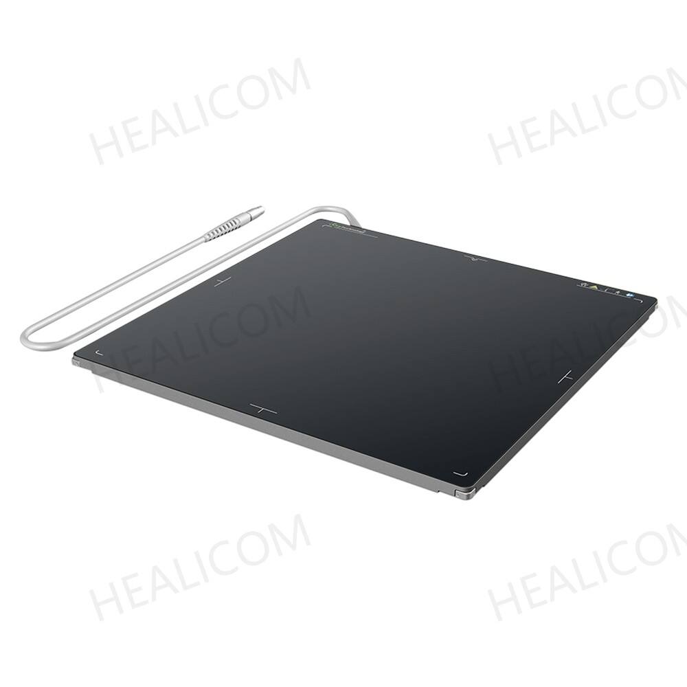
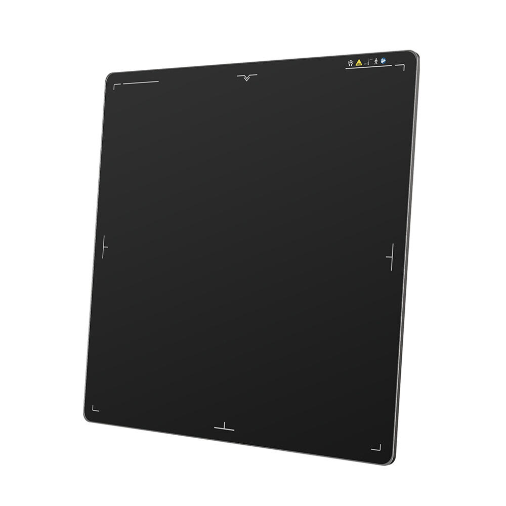
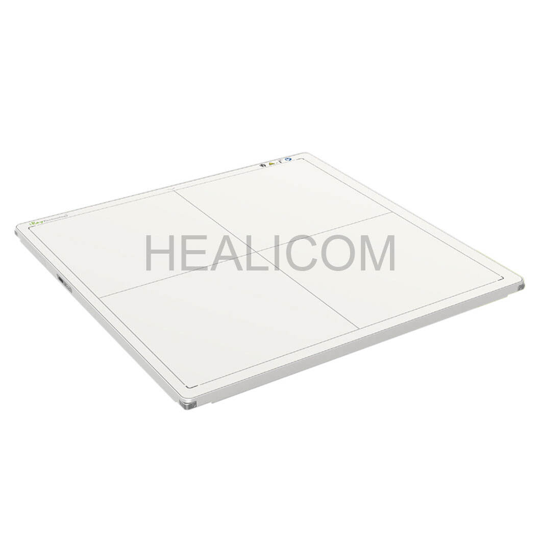
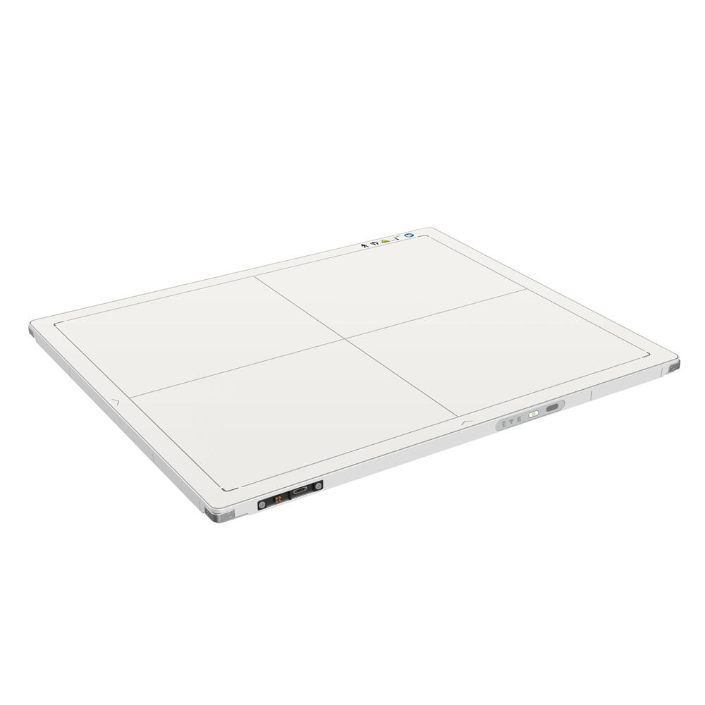
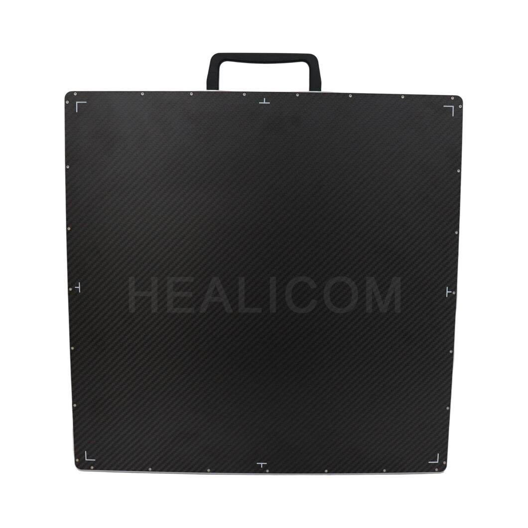
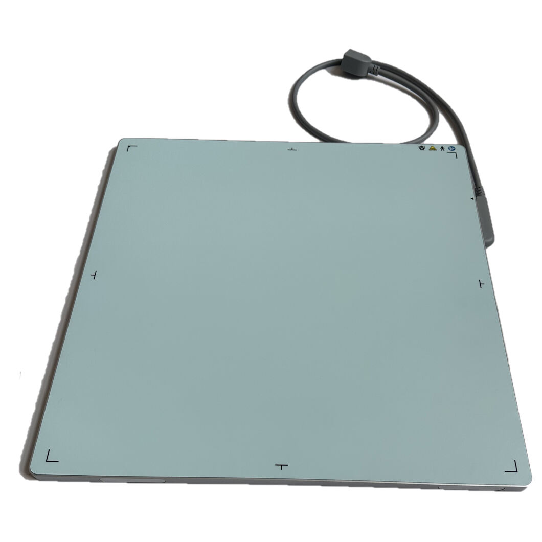
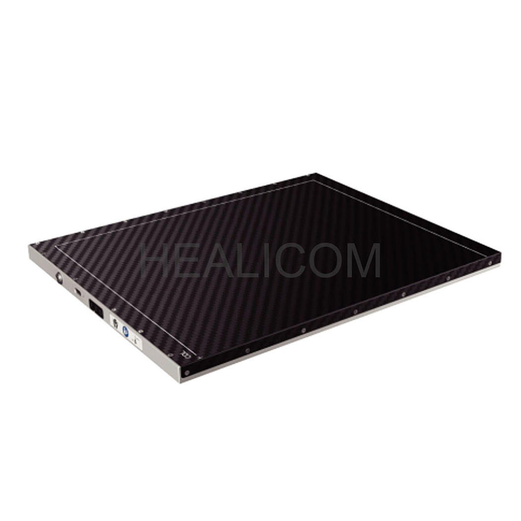
X-ray Flat Panel Detector
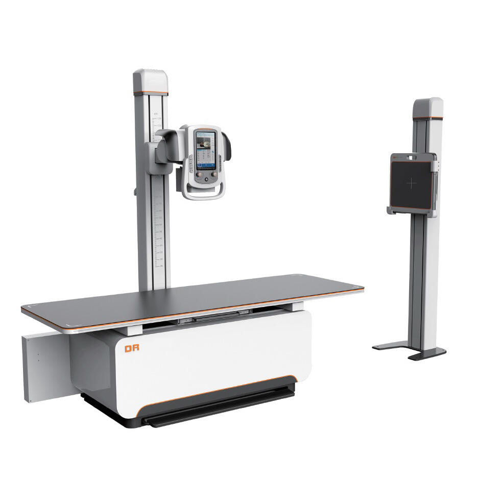
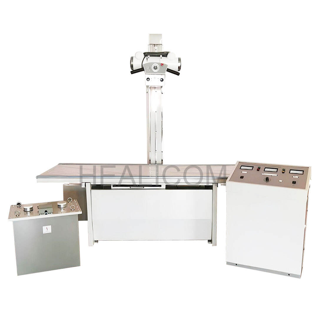
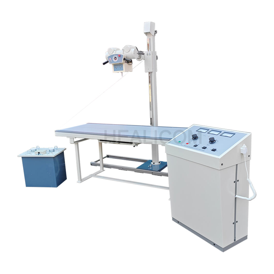
Portable X-ray System
Stationary X-ray System
Ultrasound Machine
Ultrasound Printer
Ultrasound Bone Densitometer
Ophthalmic A B Scanner
Ultrasound Probe
B,W Ultrasound Scanner
Color Doppler Ultrasound Scanner
Surgical &ICU Solution
Patient Monitor
Suction Machine
Medical Drill / Saw
Obstetrics Bed
Infusion&Syringe Pump
Electrosurgical unit
Anesthesia&Ventilator
Operation Table
Operation Lamp
Defibrillator
Medical Pendant
Medical Sterilizer
Medical Washer Disinfector
H2O2Low Temperature Plasma Sterilizer
Portable Sterilizer
Horizontal Sterilizer
Table Top Sterilizer
Vertical Sterilizer
UV Air Sterilizer
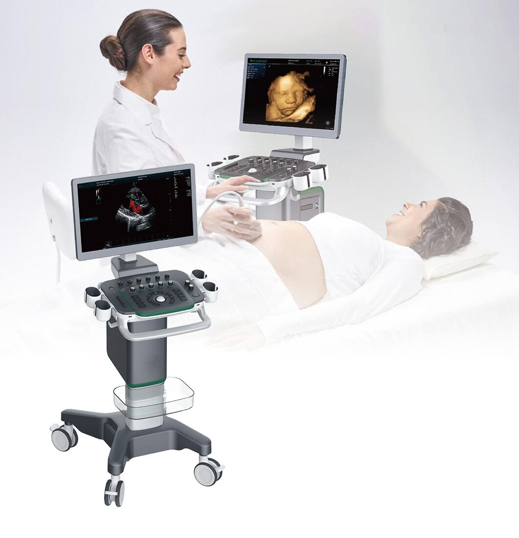
Medical User Friendly Digital Real-time Automatic Calculation Color Doppler Ultrasound
Venu 1717X
Mars 1717X
Mars 1717V
Mars 1417V
HXD-1717Y
HXD-1717W
HXD-1012
HX-300DR
HX300BZ
HX100BG
HX50R-X
PREV
1
2
3
4
...
17
NEXT
+86-13951660247
[email protected]
Scan to whatsapp :
Get a quote
Get a Free Quote
Our representative will contact you soon.
Email
Mobile/WhatsApp
Name
Company Name
Message
0/1000
Submit