EN


What Is OCT Imaging and How Does an OCT Scanner Work?
In ophthalmology, OCT usually means optical coherence tomography: a non-contact imaging method that uses light to build detailed cross-sectional views of the eye. An OCT scan is commonly associated with the retina, but the underlying technology can also be adapted for other tissue-imaging tasks. For clinical teams and equipment evaluators, the useful question is not merely whether an OCT scanner produces a sharp image. It is how the image is acquired, which workflow it supports, and what still requires professional interpretation.
This article explains the core idea behind OCT imaging, the signal path inside an OCT scanner, and the practical questions worth checking before a system is introduced into an eye-care workflow. It is educational information, not a substitute for clinical, regulatory, or engineering assessment.
What is OCT imaging?
OCT imaging is a high-resolution optical technique that measures light reflected or backscattered from different depths within tissue. In an eye examination, the result is often a layered cross-section of the retina, allowing a clinician to inspect retinal architecture rather than relying only on a surface view. That is why an OCT eye exam is frequently used alongside other parts of an ophthalmic assessment.
A helpful comparison is ultrasound, with one important difference: ultrasound uses sound echoes, whereas OCT uses light. Light can provide fine detail near the surface of scattering tissue, but it does not travel through all tissue in the same way as sound. So an OCT image should be understood as a specialized view with useful depth information—not a universal replacement for every imaging modality or clinical finding.
The basic principle: coherence tomography with light
The technical foundation of coherence tomography is low-coherence interferometry. An OCT scanner sends a broad-band, low-coherence light beam into the sample and compares returned light with light traveling along a known reference path. When those optical paths match within the source’s short coherence length, the returning signals create an interference pattern that can be detected and processed.
In practical terms, the system is looking for two pieces of information at many tiny locations: how much light comes back and from what relative depth. Differences in tissue microstructure affect the returning signal. The scanner converts those differences into a depth-resolved reflectivity profile rather than a conventional camera photograph.
How an OCT scanner builds an image
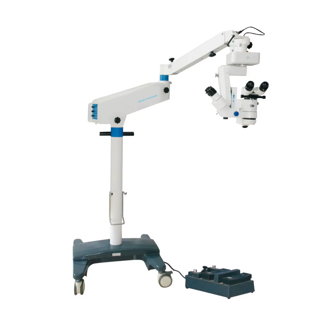
An OCT scanner contains a light source, optics that direct and focus the beam, a reference path, a detector, and software that reconstructs the signal. Exact designs vary, but the working sequence is broadly similar:
- Illuminate the target. The system directs low-power light toward the tissue. In ophthalmic use, alignment and fixation help position the scan on the intended retinal area.
- Collect returning light. Boundaries and structures within the tissue scatter or reflect part of the light back toward the instrument.
- Compare it with a reference. The returned sample signal is compared with light from the scanner’s known reference path. The interference signal carries depth-related information.
- Create an A-scan. Processing produces a one-dimensional depth profile, often called an A-scan. It represents signal intensity as a function of depth at one lateral position.
- Scan across the area. The beam moves across adjacent positions. Combining many A-scans creates a cross-sectional B-scan; repeated sections can be assembled into a volume or map.
- Review image quality and context. The operator and clinician assess centration, segmentation, artifacts, signal quality, and consistency with the patient’s wider examination.
More accurately, the image is not simply “taken” in a single instant like a standard photograph. It is reconstructed from many sampled optical signals. Motion, poor fixation, media opacity, alignment, and processing choices can therefore affect how reliably a given scan represents the target anatomy.
What an OCT eye exam can show—and what it cannot decide alone
A retinal OCT scan can help visualize layers and contour changes that may be difficult to appreciate with a surface image alone. Depending on the protocol and clinical question, a provider may use it to monitor structural changes over time, compare regions, or document findings for review. OCT angiography is a separate extension that uses repeated OCT data to derive motion-contrast information related to blood flow; it should not be confused with every standard OCT acquisition.
Still, a technically successful OCT eye exam is not the same as an automatic diagnosis. Device output must be interpreted in light of symptoms, examination findings, acquisition quality, and the intended use of the system. Automated measurements or segmentation can improve efficiency, but they should have an appropriate review process—especially when image artifacts or atypical anatomy are possible.
What to evaluate when selecting an OCT scanner
For a clinic or procurement team, start with the clinical task rather than a headline specification. A scanner that fits one workflow may be a poor match for another. The following checklist creates a more useful evaluation discussion:
- Intended applications: Which examinations, anatomical regions, and patient groups must the system support? Confirm the manufacturer’s intended use and local requirements.
- Acquisition workflow: Consider alignment assistance, fixation options, scan protocols, patient positioning, throughput, and the experience needed to obtain repeatable images.
- Image review: Check how B-scans, maps, volume data, annotations, and quality flags are displayed. Ask how the team will recognize and document artifacts.
- Data pathway: Clarify export formats, review stations, storage, connectivity, user access, and any integration requirements before deployment.
- Verification and support: Request the relevant technical documentation, training scope, maintenance pathway, and acceptance criteria. Image-quality evaluation methods matter; for example, regulatory science work has explored model-eye phantoms for assessing ophthalmic OCT image quality.
The key is to avoid treating a long feature list as proof of fit. Before comparing systems, define the required scan types, the users who will acquire and interpret images, the local data workflow, and the acceptance checks. If an automated function is available, confirm how it will be verified in the target setting rather than assuming it will work identically in every workflow.
Why image quality is a workflow issue
Good OCT imaging depends on both technology and process. A high-quality system can still yield limited information if a patient cannot maintain fixation, the scan is decentered, the ocular media reduce signal, or the operator selects an unsuitable protocol. Conversely, a thoughtful acquisition and review process makes it easier to identify when a scan should be repeated or interpreted with caution.
For that reason, an OCT implementation plan should include more than installation. It should define training responsibilities, routine quality checks, artifact escalation, data retention, and maintenance contacts. Your team should also decide who reviews exceptions and how follow-up imaging is compared over time. These are practical safeguards, not optional extras.
Key takeaways
OCT imaging uses low-coherence light and interferometric signal processing to create depth-resolved views of tissue. In eye care, an OCT scanner typically combines many depth profiles into retinal cross-sections that support examination and monitoring. The clearest way to evaluate an OCT system is to connect its scan protocols, image quality, data handling, training, and verification steps to the actual clinical workflow. A strong image is valuable, but its value ultimately depends on appropriate acquisition and expert interpretation.
 Hot News