X-ray machines work by using electromagnetic radiation to produce pictures for doctors to look at. The way they do this is pretty straightforward actually. When turned on, these machines send out controlled beams of radiation that can go right through soft tissues in our bodies but get stopped up when they hit something denser like bones or anything else that doesn't belong there. Special detectors then pick up on how much radiation gets through different parts of the body. What we see as images on film or computer screens are basically shadows created by this process. Bones show up as white areas because they block most of the radiation, while places filled with air appear dark since almost nothing stops the radiation from passing through.
Modern systems enable real-time imaging, critical for emergencies such as fractures or lung infections. Recent analyses indicate that 78% of emergency rooms now use digital x-ray systems for rapid trauma assessments, reducing diagnosis times by 40% compared to traditional methods (GlobeNewswire 2025).
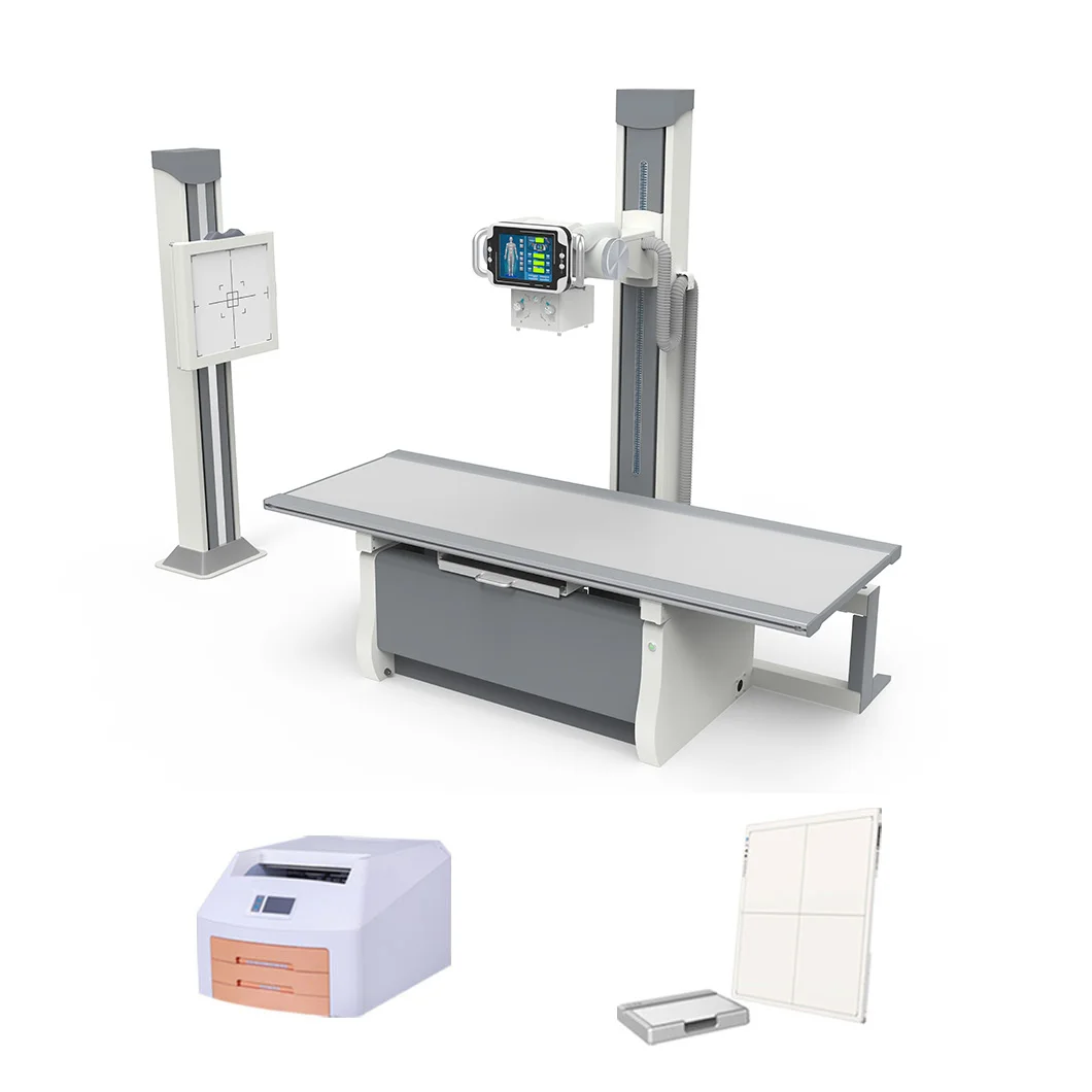
Digital Radiography, or DR for short, works with direct digital sensors that grab images right away without needing any chemicals for processing. Patients typically wait around 60% less time compared to traditional Computed Radiography methods where they have to deal with those imaging plates and separate scanning equipment. A recent paper published in Medical Physics back in 2023 found something interesting too DR actually offers about 12 percent better spatial resolution than CR does. This makes quite a difference when it comes to spotting those tricky little fractures or small lung nodules that might otherwise go unnoticed during routine exams.
Charge-coupled device (CCD) detectors are increasingly replacing older photomultiplier technologies due to their lower radiation requirements. These systems maintain diagnostic accuracy while reducing annual facility radiation costs by up to $18,000 (Journal of Diagnostic Imaging, 2024).
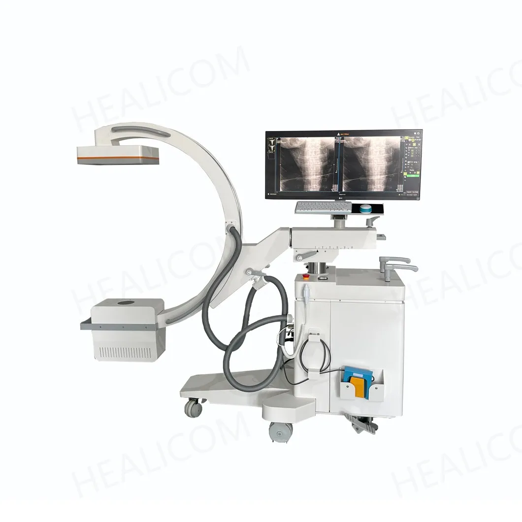
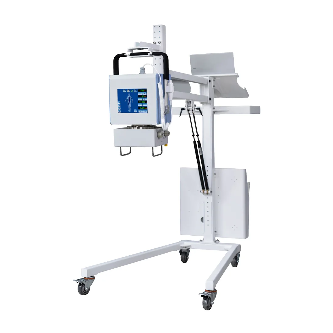
The portable X-ray machines manage around 85% image quality compared to their fixed counterparts while running on batteries for well over eight hours straight. These devices have become a must-have in intensive care units and those temporary field hospitals popping up during emergencies. Quick access to X-rays there actually cuts down trauma deaths by about 22%, according to research from EMRA back in 2023. When these units connect through IoT technology, doctors get those images sent right to them in under 90 seconds most of the time. That kind of speed really matters when making life or death decisions on the spot.
When it comes to looking at bones, X-ray machines remain one of the go to options in emergency rooms across the country. According to recent studies from the Journal of Trauma Studies published last year, around two thirds of ERs turn to plain X-rays first when assessing injuries. These machines can spot broken bones, joints out of place, and signs of wear and tear down to about a quarter millimeter detail. What's interesting is how fast they work too. The actual exposure time needed is just one thousandth of a second, which amounts to roughly what someone would normally absorb from background radiation during three regular waking hours.
Chest X-rays resolve pulmonary patterns at 0.5 lp/mm resolution, identifying early-stage pneumonia in 89% of cases. Abdominal imaging detects intestinal obstructions with 82% accuracy compared to CT scans, while using 80% less radiation. Automated exposure control in modern DR systems reduces retakes by 40% in obese patients, enhancing both safety and efficiency.
Intraoral X-rays can spot cavities down to about half a millimeter in size, which helps catch problems before they get too bad. Meanwhile, those extraoral imaging systems are pretty good at mapping out issues with the temporomandibular joint (TMJ), getting details right within just 0.6 degrees of angle. According to some research published last year in Frontiers in Dental Medicine, the latest digital detectors have reached an impressive 15 line pairs per millimeter resolution. That means tiny cracks in tooth enamel show up clearly on these images something we simply cannot see during regular checkups. Another big plus is how modern equipment adjusts exposure automatically, cutting down radiation exposure for patients by around two thirds when compared to older CR technology from years back.
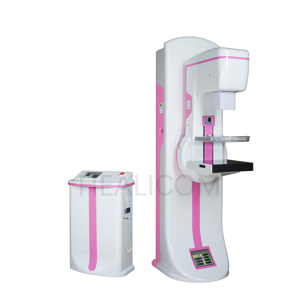
Mammograms have really changed how we approach breast cancer detection compared to just regular physical exams. The American College of Radiology says doctors can spot problems up to three years before they might otherwise be found. These tests use pretty minimal radiation actually, around 0.4 mSv each time, which is similar to what someone gets from natural background radiation over a few months. Special tools compress the breast tissue during scanning so tiny details become visible that wouldn't show up otherwise. About half of all early stage breast cancers get caught only because of routine mammograms according to recent studies. That makes a huge difference when it comes to long term outcomes for patients since catching things early often means better chances of beating the disease within five years.
When fluoroscopy works alongside contrast agents, doctors get real time images of blood vessels and can actually see how blood flows during medical procedures. Research published last year in the Journal of Vascular Interventions showed that hospitals using dynamic angiography systems cut down the time needed for stent placements by around 18 minutes compared with older static imaging methods. Cardiac cath labs now benefit from these advanced systems which can spot arterial blockages as small as 0.2 mm. To put that into perspective, imagine being able to see something as tiny as a grain of sand inside a coronary artery - that's the kind of detail these machines provide.
Modern CT scanners spin their X-ray sources around patients at roughly half a second per rotation, which transforms regular imaging data into those detailed 3D views we see on screens. The contrast for soft tissues is actually about one and a half times better than what traditional X-rays can show. Looking at the latest photon counting technology, these new CT systems are getting down to resolutions as fine as 0.1 millimeters between voxels. At the same time, they cut down radiation doses by nearly forty percent when compared to machines from just five years ago. These improvements represent something pretty significant for both diagnostic accuracy and patient safety in medical imaging.
Digital radiography is seeing major changes thanks to artificial intelligence, which speeds up image analysis by around 40 percent without compromising diagnostic accuracy. The algorithms behind AI systems have boosted their ability to spot problems in chest X rays by about 15 percent, making it easier to catch things like pneumonia or tumors at earlier stages. These smart tools definitely help streamline workflows and offer features like instant enhancements and automatic reports, but doctors still need to keep a close eye on things to avoid becoming too dependent on them. A recent study from 2025 backs this up, showing that human supervision remains critical even as technology advances.
Keeping patients safe from radiation is still at the top of everyone's list these days. New protocols have managed to cut down on patient doses by around 30 percent without messing up the image quality doctors need for diagnosis. According to research from the Ponemon Institute back in 2024, hospitals that optimize their imaging procedures can save roughly seven hundred forty thousand dollars each year just by cutting down potential legal problems. The latest technology actually uses artificial intelligence to adjust exposure levels depending on what kind of body they're scanning, which fits right in with those FDA guidelines about keeping radiation as low as reasonably possible. Most major equipment makers are now putting special software into their X-ray machines so technicians can track exactly how much radiation gets delivered during each scan.
More than three quarters of hospitals across America have made the switch from conventional radiography (CR) to digital radiography (DR) systems. The main reasons? Faster image production times, reduced day-to-day expenses, and no need for those old chemical processing labs anymore. When it comes to sharing images, cloud technology has really changed the game. Radiologists can now send scans between facilities almost instantly, which is a lifesaver for small town clinics needing expert opinions on complex cases. Market analysts are predicting big things ahead too. They think the worldwide DR market could hit around 2.5 billion dollars by the mid 2030s. Makes sense when we look at how hospitals keep pushing for quicker diagnostics while trying to cut down on waste and integrate more digital solutions into their workflows.
X-ray machines are essential in healthcare for diagnosing various conditions, from bone fractures to dental issues. They use electromagnetic radiation to create images that help doctors examine the inside of the body.
Common types include Digital Radiography (DR), Computed Radiography (CR), and portable X-ray machines. These systems differ in efficiency, image quality, and convenience.
With advancements like AI integration and adaptive protocols, modern X-ray systems reduce radiation exposure and enhance diagnostic accuracy, providing safer imaging experiences for patients.
Advanced applications include mammography for breast cancer detection, angiography for vascular imaging, and CT scans for 3D imaging of soft tissues.
AI is revolutionizing X-ray technology by improving image analysis speed and accuracy. It enables features like instant enhancements and automatic reporting, helping streamline workflows in medical imaging.
 Hot News
Whether it is business cooperation or industry exchange.