EN


How Is OCT Technology Used in Modern Ophthalmic Diagnostics?
In ophthalmology, OCT usually means optical coherence tomography: a non-invasive imaging method that uses light to build cross-sectional views of ocular tissue. An OCT eye scan does not replace the examination, patient history, or clinical judgment. It gives the eye-care team a detailed structural view that can be compared with symptoms, visual function, examination findings, and prior images.
In a busy eye-care workflow, the value of an OCT result is not simply that a scan was obtained. The useful question is whether the image is centered correctly, of adequate quality, comparable with prior studies, and interpreted in the patient’s clinical context. That is why ophthalmic imaging equipment should support a repeatable workflow rather than be judged from a feature list alone.
What an OCT Eye Scan Shows
Optical coherence tomography uses reflected light signals to create very fine, slice-like views of eye structures. In routine posterior-segment imaging, an OCT scanner machine commonly helps clinicians view the retina in layers and assess the optic nerve region. The scan can display architecture that is difficult to appreciate through a conventional surface image alone.
For the retina, OCT can help reveal the contour of the macula, the relationship between retinal layers, and the presence of structural changes such as fluid or traction when they are clinically relevant. For glaucoma assessment, OCT can provide measurements of the retinal nerve fiber layer and related optic nerve parameters. These measurements are most informative when they are reviewed alongside the examination and, where appropriate, compared over time.
An important distinction is that structural OCT and OCT angiography answer different questions. Structural OCT focuses on tissue layers and morphology. OCT angiography, often called OCTA, creates a motion-contrast view of vessels in and beneath the retina without dye. A clinical team should decide which imaging question is being asked before selecting a protocol.
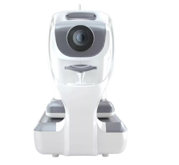
A generic editorial illustration of an OCT imaging workflow; not a clinical record or product claim.
How OCT Fits Into Diagnostic Workflows
A practical OCT workflow starts before acquisition. The operator confirms the patient, the eye, the clinical indication, and the scan protocol. During acquisition, fixation, alignment, pupil conditions, media clarity, motion, and segmentation quality can all influence the usefulness of the result. After acquisition, the clinician reviews the cross-sections rather than relying only on a color-coded summary or a single numerical output.
- Define the clinical question. Is the team assessing macular structure, optic nerve change, a treatment response, or another concern?
- Acquire a reproducible scan. Consistent centering and scan protocol make serial OCT comparisons more meaningful.
- Check image quality and artifacts. A seemingly clear report can still be affected by blink, motion, poor signal, decentration, or automated layer-boundary errors.
- Interpret in context. The OCT image is integrated with examination findings, visual acuity, symptoms, and other tests.
- Compare trends carefully. A single OCT eye scan is a snapshot; serial imaging may help clinicians evaluate whether a structural finding is stable, changing, or inconsistent with prior data.
This workflow-first approach matters because seeing a measurement on an OCT scanner machine does not automatically establish a diagnosis. In practice, the scan contributes objective structural information that qualified professionals assess with the rest of the clinical record.
Common Ophthalmic Uses of OCT
Retinal and macular assessment
In retina care, OCT is widely used to examine retinal morphology and to help document change. For example, a clinician may use an OCT image to investigate a symptom, to evaluate a suspected macular finding, or to monitor a known condition as part of an ongoing management plan. The image supports decision-making; it does not independently determine the cause of a patient’s complaint.
Glaucoma evaluation and follow-up
For glaucoma-related workups, OCT can supply structural information about the retinal nerve fiber layer and optic nerve region. The result should be read with caution. Disc appearance, intraocular pressure, visual-field testing, corneal factors, patient history, and scan quality may all affect the overall assessment. A borderline printout is not interchangeable with a confirmed diagnosis.
Anterior-segment and specialty applications
Depending on the system and intended use, OCT technology may also be applied to anterior-segment imaging or other specialty workflows. Before relying on a specific configuration, teams should verify the device’s intended use, compatible protocols, local regulatory status, and the training requirements for their setting. This is particularly relevant when comparing ophthalmic imaging equipment across sites or markets.
What to Check When Evaluating an OCT Scanner Machine
For a clinic or procurement team, the right OCT scanner machine is not necessarily the one with the longest specification list. Start with the patient populations, referral patterns, staffing model, space, data workflow, and follow-up needs. Then test whether the proposed configuration supports those tasks.
- Clinical scope: Confirm which structures, scan patterns, and applications the system is intended to support.
- Image quality and repeatability: Ask how the team will review signal quality, alignment, segmentation, and artifacts before acting on an OCT result.
- Workflow integration: Check patient positioning, scan time, operator controls, reporting, data storage, and access to prior studies.
- Training and governance: Define who acquires scans, who reviews exceptions, and how clinicians handle inadequate images.
- Service verification: Clarify maintenance, software updates, accessories, interoperability, documentation, and acceptance criteria before purchase.
In other words, do not ask only whether a system has a particular OCT feature. Ask how that feature will be verified under the clinic’s actual conditions. Image-quality assessment is a real device-performance consideration, so acceptance planning should include repeatability and quality checks rather than relying on a demonstration image.
Limits That Keep OCT Results Useful
OCT is powerful, but it has boundaries. Poor fixation, dense media opacity, motion, an off-center scan, or an algorithmic segmentation error can complicate interpretation. Normative comparisons may also be less useful when the patient does not match the population or assumptions behind the reference data. A scan must therefore be checked, not merely generated.
The same principle applies to follow-up. A change on OCT may be clinically important, but clinicians consider whether it is reproducible, whether the scan protocol matches prior testing, and whether the change agrees with other findings. This cautious approach protects against over-reading a single image and helps make optical coherence tomography a reliable part of modern diagnostic practice.
Conclusion
Modern OCT technology gives ophthalmology teams a detailed, non-invasive way to examine ocular structure and compare findings over time. Its greatest value comes from a disciplined process: choose the correct protocol, obtain a quality image, review artifacts, connect the scan with the patient’s full assessment, and document meaningful trends. When an OCT eye scan is used this way, it strengthens diagnostic conversations without pretending that technology alone can make the diagnosis.
 Hot News