EN


OCT vs Fundus Camera: Which Ophthalmic Imaging System Do You Need?
Choosing between OCT and a fundus camera starts with a practical question, not a search for one universally “better” imaging system. The two tools answer different questions. A fundus camera records an en face view of the retina and optic nerve, while an OCT system uses light to produce cross-sectional views of retinal tissue. Many practices need both perspectives; the practical decision is which clinical workflow, referral mix, documentation need, and staffing model should come first.
From a hospital procurement perspective, start with the task rather than the specification sheet. A color photograph can make a useful baseline record, support communication, and show a broad visible retinal scene. A retinal OCT scan adds depth information that helps the clinician inspect retinal layers and structural change. The choice should be verified against the work your team actually performs, not against a generic feature list.
What each imaging system shows
A fundus camera captures a photograph of the back of the eye. Standard color fundus photography commonly covers about 30 to 50 degrees of the retina and optic nerve, although the actual field depends on the system and configuration. It is especially useful when the goal is to document visible findings, compare appearance over time, or create images that are easy to review with a patient or referring clinician.
OCT, short for optical coherence tomography, is a non-invasive optical test that uses light waves to generate cross-sectional retinal pictures. Rather than replacing a photograph, an OCT scan shows a slice through tissue. This makes it useful when a clinician needs to assess retinal-layer architecture, contour, fluid-related structural changes, or optic-nerve-related measurements within the appropriate clinical context.
In day-to-day terms, fundus photography answers “what does the surface view look like?” A retinal OCT scan helps answer “what is happening through the tissue layers?” Neither image alone should be treated as a diagnosis, and the right interpretation remains a clinical responsibility.
OCT vs fundus camera: a workflow comparison
| Decision factor | OCT | Fundus camera |
|---|---|---|
| Primary view | Cross-sectional retinal or optic-nerve tissue information | En face color view of the retina and optic nerve |
| Suitable clinical task | When layer-level structural assessment is part of the clinical question | When photographic documentation, visible appearance, or broad baseline capture is the priority |
| Image-quality focus | Fixation, alignment, media clarity, scan placement, and review of signal or artifacts | Focus, illumination, field selection, media clarity, and consistent capture technique |
| Workflow output | Scans and measurement-oriented review within the clinical record | Photographs for documentation, comparison, communication, and referral records |
| Procurement question | Which scan protocols and review needs must the team support? | Which field of view and documentation workflow must the practice support? |
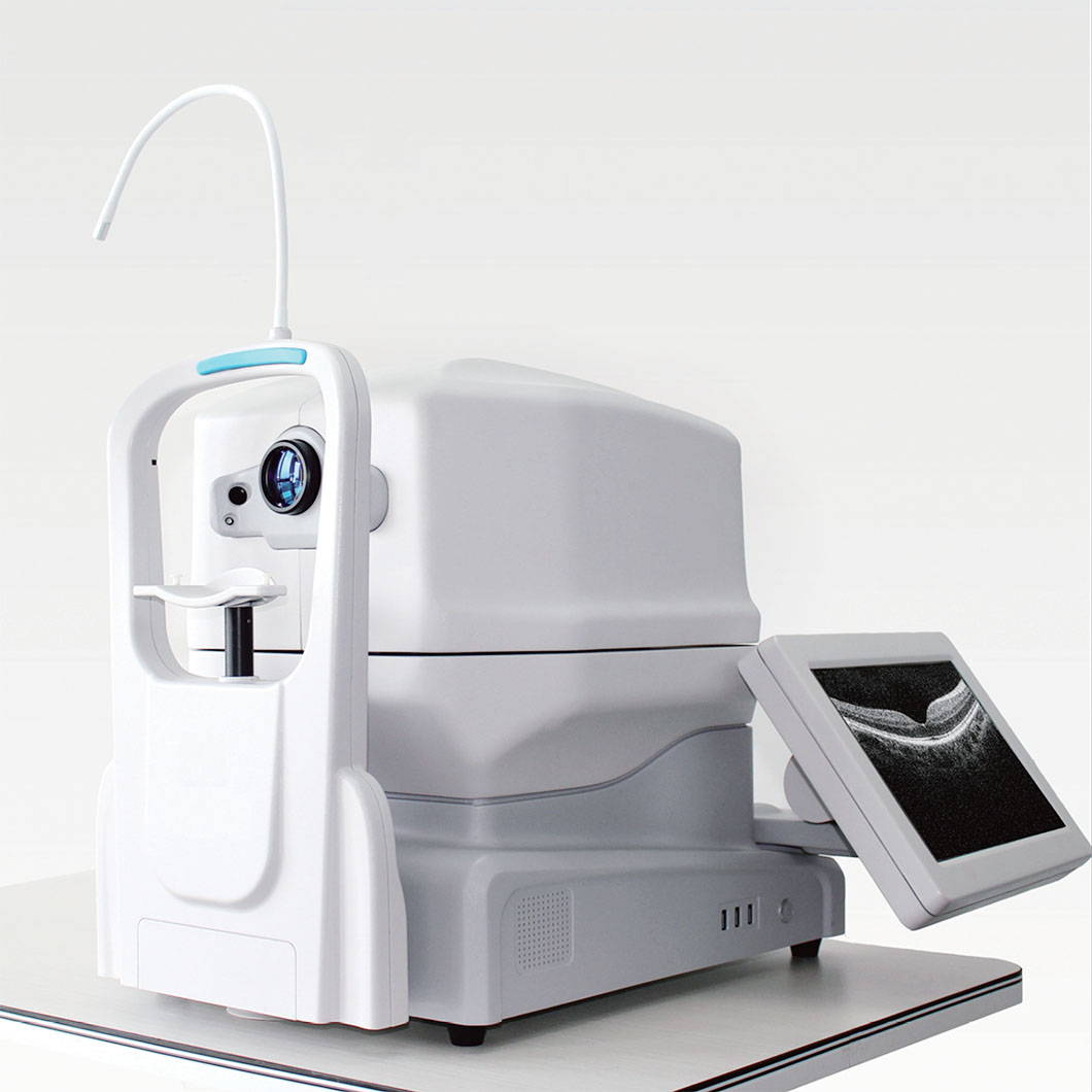 A color fundus view and a cross-sectional retinal OCT scan provide complementary information.
The comparison is deliberately conditional. A camera’s visible record can be the essential first image in one clinic, while an OCT system may be indispensable in another. If your team evaluates retinal disease, glaucoma-related optic nerve structure, or follow-up cases that require layer-level review, a suitable OCT workflow may carry more weight. If screening documentation, referral imaging, or longitudinal color records dominate, a fundus camera may be the more immediate priority.
When an OCT system should move to the top of the list
Consider prioritizing OCT when clinicians repeatedly need cross-sectional information rather than photographs alone. Typical decision signals include a retina-focused service, glaucoma assessment workflow, or a referral pattern where structural imaging changes how cases are reviewed. The key is to map the equipment to a defined task: who will acquire the scan, who will check its quality, where the result will be reviewed, and what happens when the image is not interpretable.
Do not only ask whether a device has the desired scan mode. Ask how the team will verify it under normal operating conditions. For example, a busy clinic should test the full path from patient positioning to capture, review, export, and repeat scanning. A long feature list, by itself, does not show that the system fits the appointment schedule or the people who will operate it.
When a fundus camera is the stronger first purchase
A fundus camera may be the better first investment when your highest-priority need is consistent photographic documentation. Color imaging can support baseline records, visible lesion documentation, communication with patients, and referral handoffs. It can also make it easier to compare the appearance of the posterior segment at later visits, provided capture settings and fields are used consistently.
Before selecting a camera, define whether the workflow needs standard-field images, a wider field, dilation-dependent imaging, or a particular export and record-management process. A wider field is not automatically the correct choice if the team does not have a clear use case, a review process, and training for that image type. Start with the clinical question, then confirm field, fixation support, image export, and workstation flow.
How to evaluate OCT image quality before you buy
OCT image quality is a workflow outcome, not just a number on a brochure. Because OCT records a scanning line over time rather than using a single photographic flash, fixation, motion, alignment, media opacity, and scan placement can influence the result. Demonstrations should include the patients and operating conditions that resemble your practice, not only ideal images.
- Ask the operator to acquire repeat scans and explain how motion or segmentation artifacts are identified.
- Confirm the available scan patterns match the clinical protocols your team plans to use.
- Review how the system labels laterality, stores images, and exports data to the intended record workflow.
- Check what training is included for acquisition, quality review, and basic troubleshooting.
- Define acceptance criteria before purchase, including image review, connectivity, and service response expectations.
These checks matter for all ophthalmology diagnostic devices. The value of an image depends on whether it can be acquired consistently, interpreted in context, and retrieved when the care team needs it.
Plan for complementary retinal imaging technology
In many settings, the decision is not “OCT or fundus camera forever.” It is a staged plan for complementary retinal imaging technology. The camera provides a familiar surface-view record; OCT adds sectional structure. Combining them can help a clinician correlate visible findings with underlying tissue information, but the combination only makes sense when the patient population and workflow justify it.
Procurement teams should include both clinical users and operational stakeholders. A useful evaluation meeting separates requirements into four lists: clinical questions, acquisition conditions, information handling, and lifecycle support. Then compare each candidate system against the same list. This avoids confusing a compelling demonstration with a verified fit.
A practical selection checklist
- Define the clinical question. Is the first requirement photographic documentation, cross-sectional structural review, or both?
- Map the patient journey. Identify who captures the image, who checks quality, who reviews it, and where repeat imaging fits.
- Test real-world image quality. Include fixation challenges, normal room flow, and the users who will operate the system.
- Confirm data handling. Review export, storage, identifiers, laterality, and compatibility requirements with your IT and clinical-record process.
- Compare full ownership needs. Include installation, training, maintenance, service coverage, accessories, and acceptance testing—not only the purchase price.
- Document the decision. Record why the chosen modality fits the defined workflow and which requirements remain to be verified.
Final decision: choose the question before the machine
Choose a fundus camera when a reproducible color record of the visible retina and optic nerve is the core need. Prioritize OCT when the workflow requires cross-sectional retinal or optic-nerve information that photography cannot provide on its own. If both needs are routine, plan for complementary modalities and validate the capture, review, training, and service path for each.
For buyers of ophthalmology diagnostic devices, the most useful next step is a structured demonstration checklist. Confirm the intended use, test image acquisition with actual operators, assess OCT image quality under realistic conditions, and agree on acceptance criteria before a final procurement decision. This framework supports informed equipment selection; it does not replace clinical, engineering, regulatory, or local-market assessment.
 Hot News