EN


How to Compare X-Ray Detectors and Image Acquisition Systems
Choosing an x ray machine is not only a question of generator power, room layout, or detector size. The image that users review is the output of a chain: exposure technique, detector response, acquisition workstation, processing rules, display conditions, and the quality controls around them. A practical comparison starts by defining that chain for the examinations your department actually performs.
For a procurement team, this changes the conversation with an X-ray machine supplier. Instead of asking which detector has the longest feature list, ask how the proposed detector and acquisition system support your workload, users, room constraints, and verification plan. This article provides a purchasing framework; it does not replace local clinical, radiation-safety, engineering, or regulatory review.
Start by separating the detector from the acquisition system
A detector converts the incident X-ray pattern into digital data. The acquisition system receives that data and turns it into an image that can be reviewed, processed, identified, transmitted, and archived. These components influence each other, but they should not be scored as if they were the same product.
- Detector questions: active area, portability, handling, battery and charging workflow where relevant, exposure index behavior, image-quality performance, durability expectations, and cleaning compatibility.
- Acquisition questions: workstation usability, exam worklists, patient and study identification, processing presets, repeat-analysis tools, image routing, audit trail, and connectivity with the local environment.
- System questions: generator synchronization, bucky or table integration, room workflow, display calibration, network readiness, service access, and acceptance testing.
That distinction is useful because a technically capable detector may still create friction if its acquisition workflow adds clicks, makes retakes hard to analyze, or does not fit the site’s identification and routing process.
Compare the x ray machine against real examination tasks
Before comparing specifications, list the examinations, patient-transfer patterns, throughput peaks, and operating locations that the x ray machine must support. A fixed room with high general-radiography volume has different priorities from a mobile workflow that moves between wards. For example, panel size and cable-free handling may matter more in one setting, while automated positioning, detector sharing, or rapid room turnover may matter more in another.
Build a simple task matrix with the radiographer, biomedical or clinical engineering representative, IT contact, and procurement lead. For every task, document the patient position, detector placement, expected image review point, required routing destination, and what would make a repeat exposure avoidable. This brings the comparison back to work rather than brochure language.
Read detector specifications in context
Pixel pitch, spatial resolution, detector size, and dose-related image-quality claims can be useful inputs, but none is a stand-alone purchasing conclusion. Detective quantum efficiency (DQE), for instance, is commonly used to describe how an imaging device preserves signal-to-noise information. IEC 62220 provides a method for determining DQE for digital X-ray imaging devices. When discussing a DQE value, ask the supplier to clarify the test method, beam quality, dose conditions, image-processing state, detector size, and whether the compared values describe like-for-like configurations.
It is also worth checking what happens at the edges of normal work: pediatric or larger-body-part techniques, grid and non-grid exams, portable positioning, time between exposures, battery changes, detector warm-up behavior, and recovery after a communication interruption. Those observations may reveal more about daily usability than a single headline number.
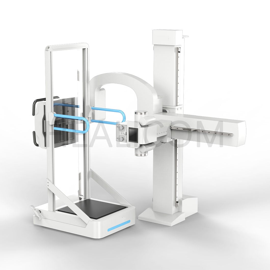
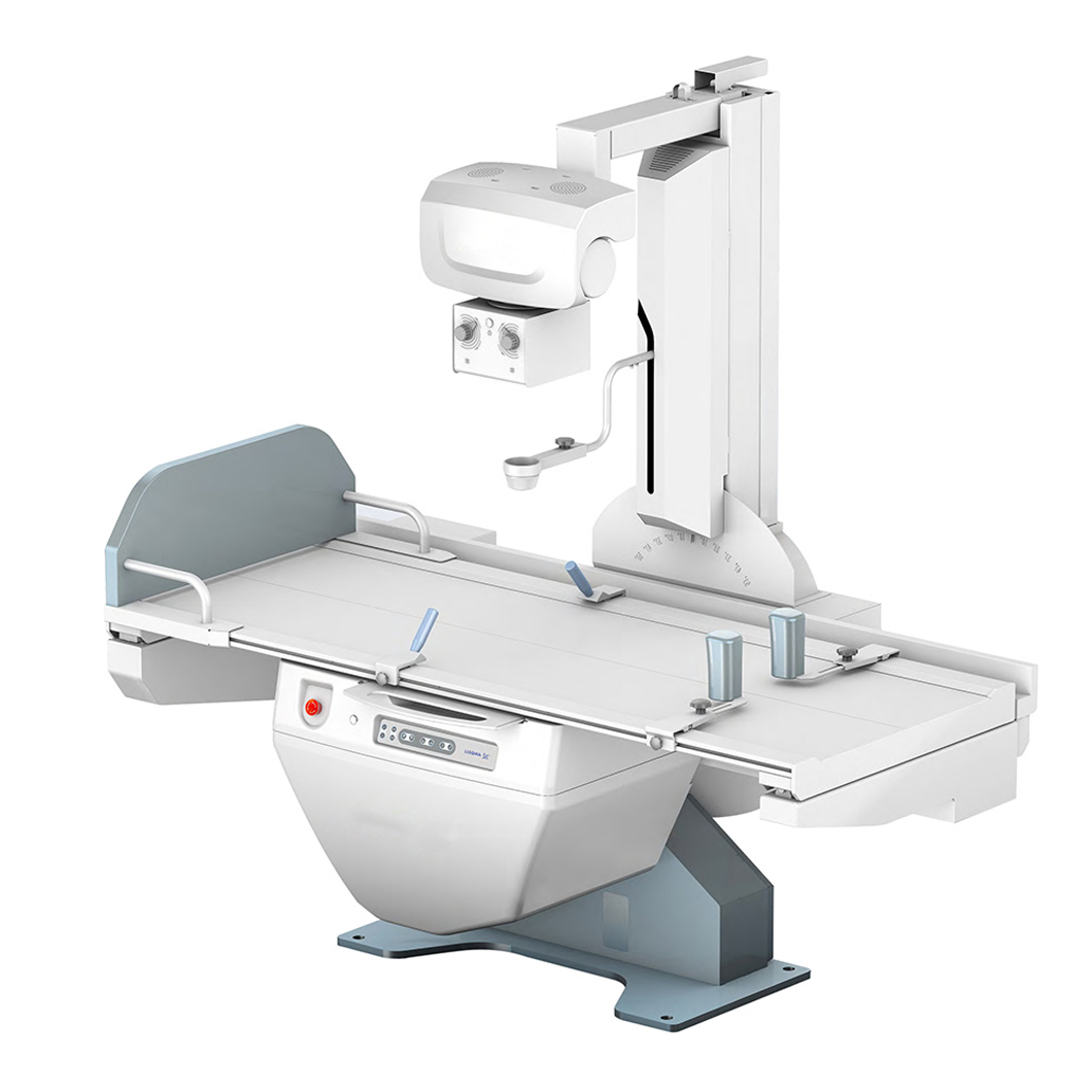 Detector and acquisition workflow should be assessed as one imaging chain.
Evaluate image acquisition as a workflow, not a screen
Image acquisition software should help users complete the examination accurately and consistently. During a demonstration, follow a representative study from worklist selection to image review and routing. Observe how the system handles patient identity, body-part and projection selection, laterality, annotations, exposure feedback, rejected-image reasons, and corrections. The relevant question is not whether a feature exists; it is whether the team can use it reliably within the intended workflow.
Ask to see how processing presets are governed. A preset that produces a pleasing image on a demonstration monitor may require different local review after the target room, display, protocol, and clinical task are defined. Procurement documentation should therefore state which settings can be configured, who can change them, how changes are recorded, and how the team will validate them during commissioning.
Make radiography quality assurance part of the comparison
Radiography quality assurance should begin before the purchase order is finalized. Current IEC 61223-3-8 guidance addresses acceptance and constancy testing of imaging performance and related quality-control parameters for radiography and radioscopy, describing the imaging chain from acquisition through processing and display. The point for buyers is practical: agree on the evidence and test responsibilities before installation, not after a problem appears.
A useful acceptance plan can include:
- Agreed configuration identifiers for the detector, acquisition workstation, software version, and room components.
- Named test phantoms, baseline image-quality checks, exposure-index review, display conditions, and repeat-analysis review appropriate to local procedures.
- Defined responsibilities for supplier staff, the responsible organization, physicists or qualified local experts, and service personnel.
- A change-control method for software, processing presets, detector replacement, and network changes.
These items are not a universal test protocol. They are the questions that keep a comparison focused on verifiable performance and local accountability.
Plan X-ray room installation before selecting the final configuration
X-ray room installation affects both the equipment choice and the implementation schedule. Confirm room dimensions, structural and shielding requirements, power, grounding, cable paths, network provisions, detector storage and charging locations, patient movement routes, and display placement. If a mobile workflow is under consideration, also map storage, charging, infection-control procedures, and handoff points between teams.
Ask each bidder to state the assumptions behind its installation scope. A quotation can appear comparable while excluding different site work, interfaces, accessories, training, or commissioning activities. Place those assumptions in one side-by-side schedule so the comparison does not turn into a price comparison between unlike configurations.
Use a procurement scorecard that shows trade-offs
| Decision area | What to compare | Evidence to request |
|---|---|---|
| Clinical tasks | Exam mix, patient handling, location, throughput | Workflow walkthrough and task matrix |
| Detector fit | Size, handling, image-quality conditions, maintenance | Configuration sheet and test-condition notes |
| Acquisition workflow | Identification, presets, retake review, routing | Demonstration using representative scenarios |
| Integration | Interfaces, network, storage, display behavior | Connectivity responsibility matrix |
| Quality controls | Acceptance baseline, constancy checks, change control | Quality-control manual and commissioning plan |
| Project delivery | Site work, training, service, handover | Installation schedule and responsibility list |
Questions to ask an X-ray machine supplier
- Which detector and acquisition-software versions are included, and how are their identifiers recorded?
- What conditions apply to the presented image-quality measurements?
- Can the proposed workflow be demonstrated using our typical examinations and routing steps?
- Which connectivity, room-preparation, and commissioning responsibilities remain with our organization?
- What training covers radiographers, technical support personnel, and administrators?
- How are processing changes, software updates, and detector replacements documented and revalidated?
For medical imaging procurement, a clear answer is often more valuable than a broad claim. A supplier should be able to define the configuration, assumptions, handover evidence, and items requiring local confirmation.
Conclusion: choose the chain, then verify it
A sound x ray machine comparison considers detector capability, acquisition workflow, room readiness, integration, and ongoing quality control together. Start with the work your team must complete, compare equivalent configurations, and write acceptance evidence into the project plan. That approach helps procurement teams make a transparent decision while leaving clinical and regulatory determinations to the qualified people responsible for them.
 Hot News