EN


What Quality Assurance Checks Are Needed for C-Arm Equipment?
A dependable carm quality assurance program checks more than whether an image appears on screen. The team must also know whether the unit moves safely, produces a consistent image at an appropriate radiation output, displays the right information and can be taken out of service when a fault occurs. These checks link daily use with scheduled technical testing and a documented response to abnormal results.
For biomedical engineers, radiographers, medical physicists and operating-room teams, the useful question is not simply “Does the carm work?” It is “What was checked, against which baseline, by whom, and what happens if the result changes?” This guide outlines a practical framework; it does not replace the manufacturer's instructions, a medical physicist's assessment or local radiation rules.
Start with acceptance testing and a baseline
Before clinical use, qualified personnel should verify that the carm delivered matches the agreed configuration and that installation, labeling, safety features and documentation are in order. A medical physicist or other locally authorized specialist should carry out or oversee the required acceptance measurements. Record the model, serial number, software version, detector, test conditions, results and any outstanding deficiencies.
Establish baseline images and measurements for the C-arm imaging system under reproducible settings. Depending on the equipment and local protocol, those may include image uniformity, spatial resolution, low-contrast visibility, radiation output and automatic exposure or brightness control performance. A baseline is useful only if later tests use comparable geometry, phantoms and settings. Agree beforehand on action limits and who can authorize return to use.
After a major repair, detector replacement, calibration or software change, a carm may need targeted testing before it returns to clinical service. The scope depends on what changed and on the manufacturer's instructions and local procedures; a routine operator check is not a substitute for this assessment.
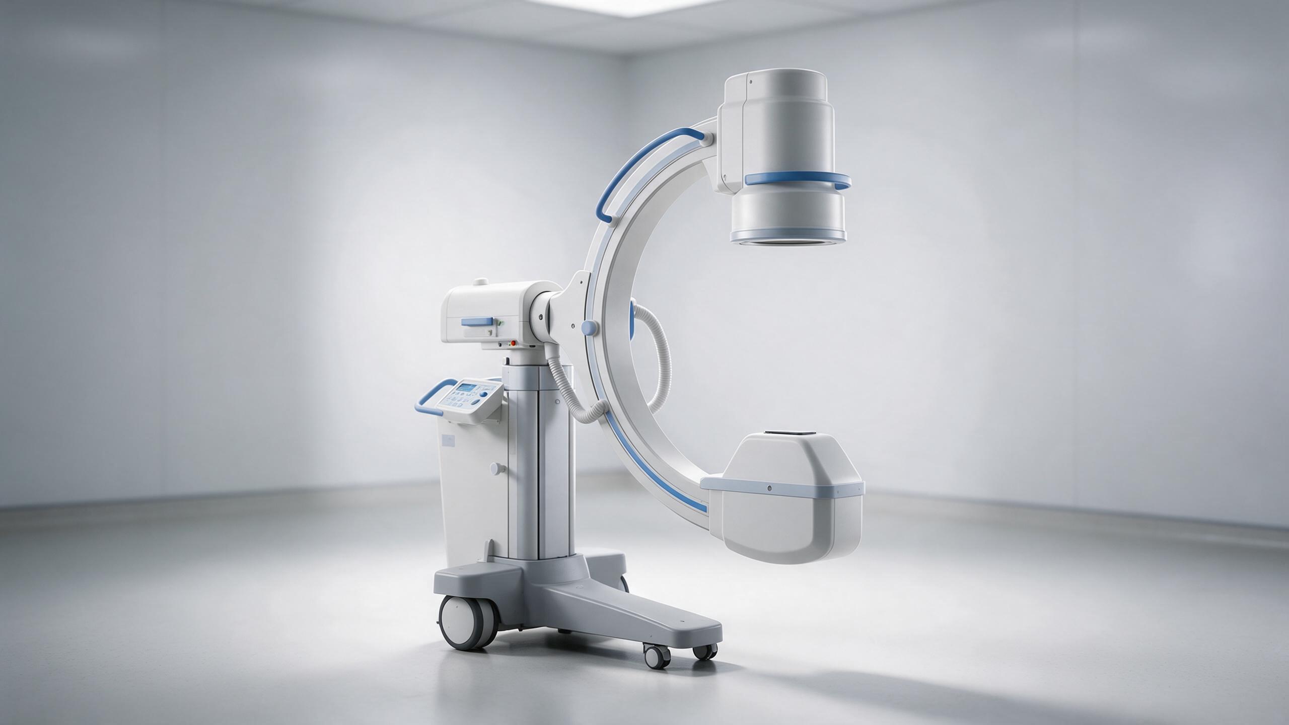
Check the carm before use
A trained operator can complete a short inspection before the first case or at another interval set by the facility. Do not expose a patient merely to perform a test. Any test exposure must follow the approved local procedure, use appropriate shielding and be performed by authorized personnel.
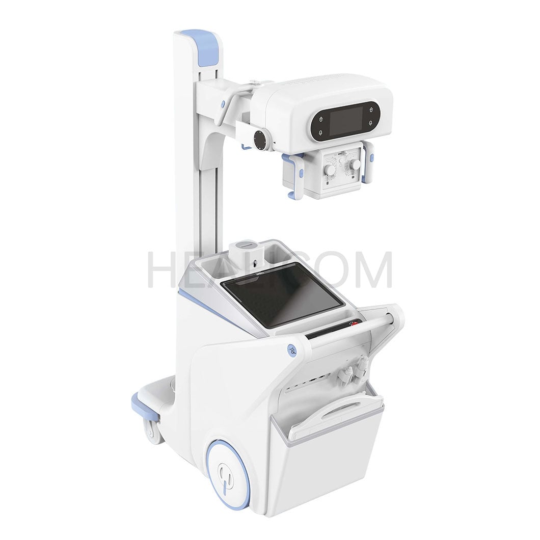
- Mechanical condition: Inspect the carm frame, covers, cables and connectors for damage. Test wheels, brakes, locks and permitted movements; confirm that the arm holds position and does not drift or collide with nearby equipment.
- Electrical and controls: Check the power lead and accessible controls. Confirm that the emergency stop, footswitch and X-ray activation controls operate as specified, without unintended activation. Do not open electrical housings.
- Displays and warnings: Check monitor visibility, status messages, exposure indicators and audible signals. Confirm that the display and last-image-hold function work when applicable to the model.
- Cleanliness and accessories: Inspect surfaces and accessories for damage or contamination. Use cleaning agents and methods approved for the equipment and the facility's infection-control procedures.
Keep a dated log for each carm, including the outcome, initials and any escalation. An image artifact, loose cable or failed brake is not a cosmetic issue to hide in a checklist. Stop use when a safety feature fails, label the unit and notify the appropriate service and radiation-safety contacts under the local policy.
Assess image quality against the carm baseline
For a mobile fluoroscopy machine, the clinical image and the test image answer different questions. The clinical team should report changes in visibility during normal work, while a qualified tester uses a suitable phantom and a repeatable protocol to investigate performance. Comparing images taken with different modes or geometry can create a false impression of change.
The IAEA's diagnostic radiology quality-control handbook describes checks for low-contrast detail, spatial resolution, uniformity, distortion and artifacts. It includes comparison with baseline performance. The local medical physicist should select the phantom, acceptance criteria and testing interval for the specific carm; the handbook's example frequencies are not a universal schedule.
Look for new bands, dead areas, unusual noise or uneven brightness. Confirm that the displayed orientation and any image markers agree with the acquisition. If the image changes, document the imaging mode, settings and the affected detector area. A service engineer can then investigate, and an authorized specialist can decide which repeat tests are needed before release.
Verify radiation output and dose information
The carm quality program should address both image quality and radiation output. A medical physicist can measure output, evaluate automatic control response and check the relationship between image quality and dose using calibrated instruments and a defined setup. Increased output with little improvement in visibility deserves investigation; it is not something the operator should correct by guessing settings.
Where the C-arm imaging system provides dose indicators, check that the displays are readable and that staff understand their meaning. Reference air kerma and kerma-area product are useful indicators, but a displayed reference air kerma is not automatically the patient's peak skin dose. Local protocols should define how dose information is reviewed and documented, especially for lengthy or repeated procedures.
US requirements in 21 CFR 1020.32 address features such as irradiation-time information, last-image hold and dose displays for covered fluoroscopic equipment, with provisions that depend on manufacture date and equipment type. Other jurisdictions may differ. Confirm the applicable version, local rules and model-specific functions rather than assuming that every carm has identical displays.
Review workflow, safety and data transfer
Quality assurance also covers the way surgical imaging equipment is used. Confirm that trained staff can select the intended imaging mode, position the detector and use collimation and available dose-management features according to the clinical protocol. Review shielding, staff positioning and personal monitoring with the radiation-safety team. The goal is an image fit for the clinical task with exposure managed appropriately, not the darkest or brightest image possible.
For intraoperative X-ray, verify patient identification, image labeling and transfer to the destination used by the facility. Test connectivity and retrieval using an approved test workflow, not real patient information in an uncontrolled environment. A successful image on the carm screen does not by itself confirm that the correct study was stored in the correct record.
Inspect the monitor in its actual working position. Reflections, an obscured display or difficult access to the footswitch can affect use even when the carm passes a technical test. Record issues with the clinical team and refer them to the person responsible for the room layout or training.
Set a schedule and act on failed checks
Use a written plan that assigns pre-use checks to trained operators, scheduled service to biomedical engineering or an authorized provider, and radiation and image-performance tests to the medical physicist or other qualified person. Set intervals using the manufacturer's instructions, local regulations, facility policy, usage and risk assessment. Document any required checks after repair or a reported incident.
Each carm record should include the test, date, responsible person, method, baseline or limit, result, fault report, corrective action and authorization to resume use. Trend comparable results over time rather than waiting for a single obvious failure. If a test is overdue or an action limit is exceeded, follow the facility's escalation and equipment withdrawal procedure.
A mobile fluoroscopy machine is ready for work only when its condition, image performance, radiation safeguards and records support the intended use. Review the quality plan with the people who operate and maintain the unit, and ask the supplier for the model-specific manuals, service requirements and acceptance-test documentation before planning the next inspection.
 Hot News