Digital radiography X-ray machines achieve radiation dose reduction through fundamental improvements in detector physics. Unlike legacy systems that required high exposures to compensate for inefficient photon capture, modern detectors convert over 90% of X-ray photons into usable signals via two key advancements.
Detectors with DQE scores above 75% at 60 kVp enable 30–50% lower patient doses while maintaining diagnostic clarity. This efficiency arises from optimized charge collection in materials like amorphous selenium, which demonstrates 95% quantum efficiency across diagnostic energy ranges according to quantum photonics research.
Amorphous selenium’s direct conversion architecture eliminates light-scattering losses inherent in traditional scintillator-based systems. Its uniform structure enables precise 1:1 photon-to-electron conversion, unlike indirect detectors that lose 15–20% of signal through fiber optic tapers.
A 2023 multicenter trial published in the Journal of Medical Imaging demonstrated a 62% lower effective dose in pediatric chest exams using selenium-based detectors compared to CR systems. Image quality remained equivalent (4.1/5 vs. 4.0/5) despite reduced exposure.
Current R&D focuses on graphene-oxide hybrid detectors showing 120% higher DQE than silicon in prototype testing. Photon-counting spectral detectors now entering clinical trials promise an additional 40% dose reduction through energy-specific photon sorting.
Having images available right away cuts down on retakes and reduces unnecessary exposure for patients. Digital radiography or DR systems get rid of those annoying film processing waits because they show real time previews of the images. This lets techs check if everything is positioned correctly and whether the exposure settings were good enough. According to a study published in Radiology Practice back in 2022, hospitals that switched to direct digital capture saw their repeat scan rates drop between 33% and almost half compared to older CR systems. That means less radiation for patients overall since there's no need to do extra scans when the first one works out.
Workflow Benefits of Wireless Detectors and Real-Time Review
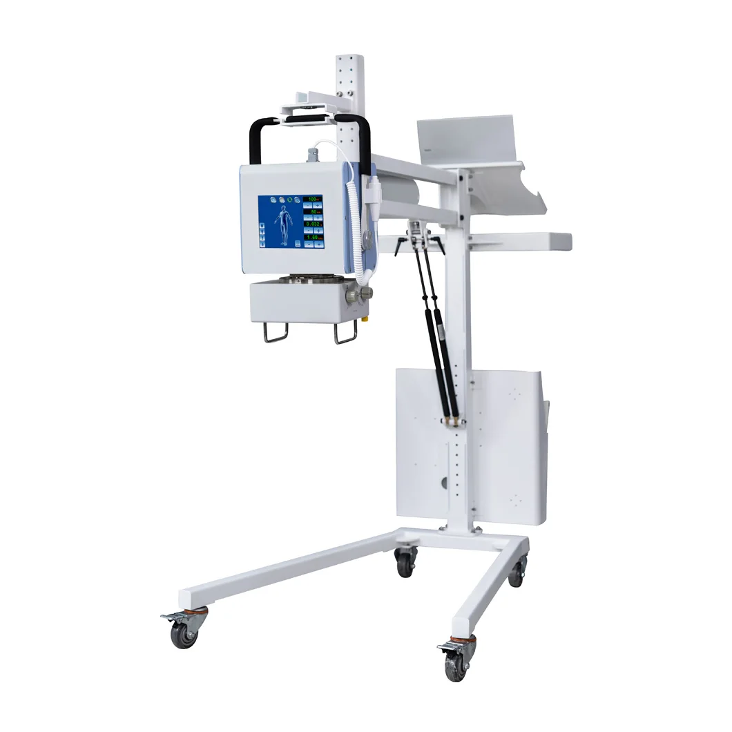
Portable DR detectors transmit images wirelessly within 15–20 seconds, enabling clinicians to identify suboptimal exams before the patient leaves the table. This prevents recall visits caused by post-processing discovery of errors—a frequent issue with CR.
Case Study: Faster Throughput in Emergency Departments with Fewer Retakes
A level 1 trauma center reduced unnecessary pelvic X-rays by 41% (p<0.001) after deploying wireless DR detectors with edge-enhancement software. Real-time collaboration cut average exam times from 12.3 to 8.7 minutes, maintaining diagnostic accuracy (J. Emerg. Med. 2023).
Portable and wireless digital radiography systems are becoming a big part of everyday clinical practice these days. Many hospitals have started using these mobile DR units equipped with lighter panels, which actually cut down on positioning mistakes during bedside imaging. A recent study across multiple sites showed this approach reduced errors by around 22%. Looking ahead, most experts predict that nearly nine out of ten new X-ray setups will go completely wireless by 2026 according to IMV Medical's latest report from last year. This shift is happening fast mainly because regulations requiring lower radiation doses are getting stricter all over the healthcare industry.
Modern digital radiography X-ray machines use automatic exposure control (AEC) systems that dynamically adjust radiation output based on real-time anatomical analysis. These systems minimize overexposure by responding to tissue density variations and patient-specific factors such as BMI or age.
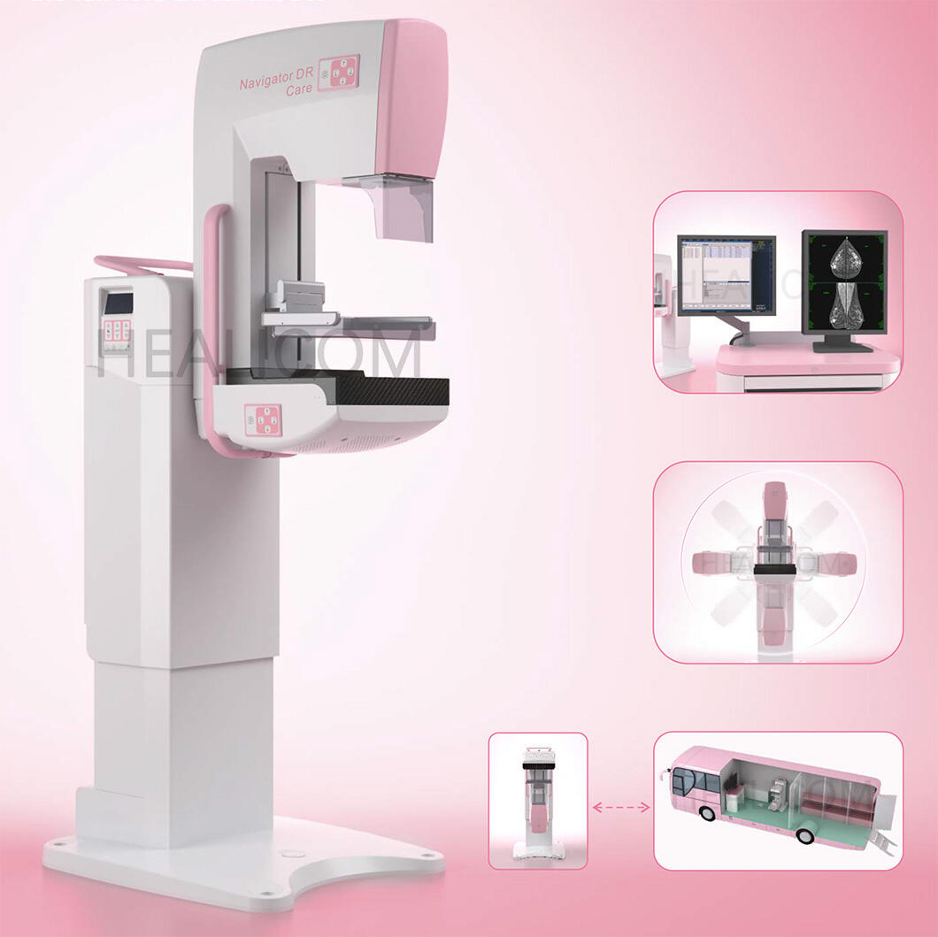
AEC sensors detect tissue composition differences through iterative exposure assessment, automatically modulating beam intensity. For instance, thoracic imaging requires 22% less radiation for pediatric patients than adults due to thinner chest walls (IAEA 2023 guidelines). This precision protects radiosensitive tissues like breast tissue during chest X-rays.
Real-time ionization chambers measure radiation reaching the detector, enabling closed-loop adjustments. If initial exposure achieves sufficient contrast, the system terminates the beam early—reducing doses by 15–30% in abdominal studies compared to fixed protocols.
A 2023 multi-center analysis demonstrated AEC systems reduced dose variability by 40% across 27 healthcare facilities. In lumbar spine imaging, median doses decreased from 4.2 mGy to 2.8 mGy without sacrificing diagnostic accuracy.
Some radiologists report gradual annual dose increases of 5–8% when operators rely too heavily on automation. Regular phantom testing and AEC recalibration every six months mitigate this risk by ensuring consistent system sensitivity.
Leading institutions implement protocol-specific AEC profiles, with studies showing 29% lower knee imaging doses when using pediatric versus adult settings. Daily quality assurance checks confirm detector response consistency across all anatomical programs.
 Hot News
Whether it is business cooperation or industry exchange.